 100% FREE ACCESS
12th Standard Syllabus & Materials
12th Standard
TN 12th Maths Applications of Matrices and Determinants Sample Question Papers Study Material - QB365 Set A
NEW12th Standard
TN 12th Computer Applications மின்னணு தரவு பரிமாற்றம் Sample Question Papers Study Material - QB365 Set A
NEW12th Standard
TN 12th Computer Applications மின் - வணிக பாதுகாப்பு அமைப்புகள் Sample Question Papers Study Material - QB365 Set A
NEW12th Standard
TN 12th Computer Applications மின்னணு செலுத்தல் முறைகள் Sample Question Papers Study Material - QB365 Set A
NEW12th Standard
TN 12th Computer Applications மின் - வணிகம் Sample Question Papers Study Material - QB365 Set A
NEW12th Standard
TN 12th Computer Applications திறந்த மூல கருத்துருக்கள் Sample Question Papers Study Material - QB365 Set A
Published on: 26/08/2022
QB365 provides a detailed and simple solution for every Possible Creative Questions in Class 12 Biology Subject - Zoology - Human Reproduction, English Medium. It will help Students to get more practice questions, Students can Practice these question papers in addition to score best marks.
Download Tamil Nadu 12th Standard Biology question papers, model tests, one-mark questions, important questions, and public exam papers in PDF format. Free study materials and answer keys for TN State Board students.
Questions + Answers key
Take MCQ Biology Test1.
(a) In which part of the fallopian tube does fertilization occur in humans? Describe the development of a fertilized ovum up to implantation.
(b) How is polyspermy prevented in humans?
2.
(a) Name the hormones secreted and write their functions.
(i) By corpus luteum and placenta (any two)
(ii) During follicular phase and parturition.
(b) Name the stages in a human female where:
(i) Corpus luteum and placenta co-exist
(ii) Corpus luteum temporarily ceases to exist
3.
(a) Arrange the following hormones in sequence of their secretion in a pregnant woman.
(b) Mention their source and the function they perform: hCG; LH; FSH; Relaxin
4.
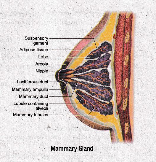
With a suitable diagram, describe the organisation of Mammary glands.
5.
The zygote passes through several developmental stages till implantation. Describe each stage briefly
6.
Name the three primary germ layers of embryo. Also, mention any three organs or organ systems developing from each layer.
7.
Write a note on embryonic membranes
8.
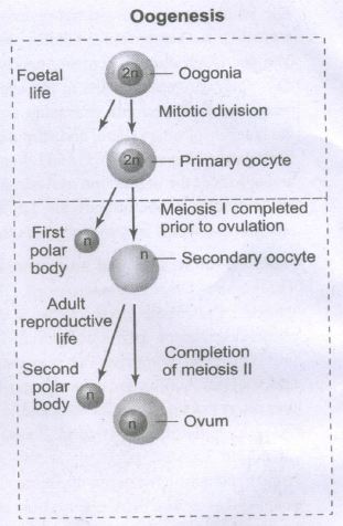
Explain the process of oogenesis.
9.
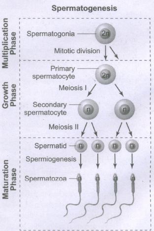
Describe the spermatogenesis with diagram.
10.
Explain the structure and function of mammary glands.
11.
Describe the structure of human ovary.
12.
Write a note a extra embryonic membranes.
13.
Explain the process of fertilization in human beings.
14.
Describe the structure of a sperm. (or) Describe the structure of human sperm with a neat labelled diagram.
15.
Differentiate Spermatogenesis and oogenesis.
1.
(a) Fertilization occurs in the ampullary or ampullary isthemic junction.
(i) Cleavage occurs in the zygote to form 2,4, 8,16 blastomeres.
(ii) 8-16 blastomere stage is called morula.
(iii) The morula continues to divide and transforms into blastocyst.
(iv) The blastomeres in the blastocyst are arranged into an outer trophoblast which gets attached to the endometrium by zona pellucida.
(v) The inner cell mass gets differentiated into an embryo.
(vi) The uterine cells divides rapidly and covers the blastocyst leading to implantation.
(b) When a sperm comes in contact with the Zona pellucida layer of the ovum, it induces changes in the membrane to block further the entry of additional sperms.
2.
(a) (i) Corpus luteum secretes progesterone which is essential for maintenance of endometrium.
(ii) Placenta secretes hCG which stimulates and maintains corpus luteum for secretion of progesterone.
(iii) hpL is helpful in supplying energy to the growing foetus.
(iv) Estrogen supports foetal growth and brings about metabolic changes in mother.
(v) Progesterone helps in maintenance of pregnancy.
(b) (ii) During follicular phase:
FSH stimulates follicular development
During Parturition:
Oxytocin causes stronger contraction of smooth uterine muscles.
(i) They co-exist during pregnancy or gestation.
(ii) It happens during menstruation.
3.
(a) FSH, LH, hCG, Relaxin
(b)
| Hormone | Source | Function |
| a) FSH | Anterior Pituitary | Stimulates follicular development |
| b) LH | Anterior Pituitary | Release of ovum from graafian follicles (ovulation) |
| c) hCG | Placenta | Supports foetal growth |
| d) Relaxin | Placenta | Softens pubic symphysis during later stages of pregnancy |
4.
The description of mammary gland is as follows:
(i) The mammary glands are paired structures (breasts) that contain undeveloped duct system and adipose tissue. (Fat containing cells)
(ii) During pregnancy, in response to oestrogen and progesterone, a glandular system develops for milk productions (lactation).
(iii) Glandular tissue of each breast develops mammary lobes containing clusters of cells called alveoli.
(iv) The cells of alveoli secrete milk, which is stored in the cavities (lumens) of alveoli.
(v) The alveoli open into mammary tubules. The tubules of each lobe join to form a mammary duct opens exterior as nipple or teat.
(vi) Several mammary ducts join to form a wider mammary ampulla which is connected to lactiferous duct through which milk is sucked out by the baby.
5.
The zygote passes through the following stages till implantations.
(i) The embryo with 8-16 blastomeres is called a morula formed from one celled zygote.
(ii) The morula continues to divide mitotically and transform into blastocyst as it moves further inio uterus.
(iii) The blastomeres in the blastocyst are arranged into a surface layer called trophoblast and a cluster of interior cells attached to trophoblast are called the inner cell mass.
(iv) The trophoblast layer then gets attached to the endometrium and inner mass cells gets differentiated as embryo.
(v) After attachment, the uterine cells divide rapidly to cover the blastocyst.
(vi) The blastocyst becomes embedded in the uterine endometrium. This is called implantation.
6.
| Germ Layers | Organ (or) Organ Systems | |
|---|---|---|
| a. | Ectoderms | Central nervous system, peripheral nervous system, and mammary gland. |
| b. | Mesoderm | Cartilage and bones, muscles, urinogenital organs |
| c | Endoderm | Epithelium of respiratory tract, liver, thyroid gland. |
7.
The extra embryonic membranes include amnion, yolk sac, allantois, and chorion. They protect the embryo from desiccation, mechanical shock, absorption of nutrients, gaseous exchange and placental formation.
| Embryonic membrane | Function | |
| a. | Amnion | Innermost two-layered membrane with amniotic fluid providing buoyancy to embryo from injury regulates temperature and acts as a medium for fetal movement. |
| b. | Chorion | Outermost layer covering and protecting the embryo. |
| c. | Allantois | Forms an outpocketing of embryonic tissue at the tail of yolk sac which forms umbilical cord that links embryo to placenta and finally becomes the part of urinary bladder |
| d. | Yolk sac | Forms a part of gut and acts a source of earliest blood cells and blood vessels |
8.
Oogenesis is the process of development of the female gamete or ovum or egg in the ovaries. During foetal development, certain cells in the germinal epithelium of the foetal ovary divide by mitosis and produce millions of egg mother cells or oogonia. No more oogonia are formed or added after birth. The oogonial cells start dividing and enter into Prophase- I of meiotic division-I to form the primary oocytes which are temporarily arrested at this stage. The primary oocytes then get surrounded by a single layer of granulosa cells to form the primordial or primary follicles. A large number of follicles degenerate during the period from birth to puberty, so at puberty, only 60,000 to 80,000 follicles are left in each ovary. The primary follicle gets surrounded by many layers of granulosa cells and a new theca layer to form the secondary follicle. A fluid filled space, the antrum develops in the follicle and gets transformed into a tertiary follicle. The theca layer gets organized into an inner theca intema and an outer theca extema. At this time, the primary oocyte within the tertiary follicle grows in size and completes its first meiotic division and forms the secondary oocyte. It is an unequal division resulting in the formation of a large haploid secondary oocyte and a first polar body. The first polar body disintegrates. During fertilization, the secondary oocyte undergoes second meiotic division and produces a large cell, the ovum, and a second polar body. The second polar body also degenerates. The tertiary follicle eventually becomes a mature follicle or Graafian follicle. If fertilization does not take place, the second meiotic division is never completed and the egg disintegrates. At the end of gametogenesis in females, each primary oocyte gives rise to only one haploid ovum.
9.
Spermatogenesis is the sequence of events in the seminiferous tubules of the testes that produce the male gametes, the sperms. During development, the primordial germ cells migrate into the testes and become immature germ cells called sperm mother cells or spermatogonia in the inner surfaces of the seminiferous tubules. The spermatogonia begin to undergo mitotic division at puberty and continue throughout life. In the first stage of spermatogenesis, the spermatogonia migrate among Sertoli cells towards the central lumen of the seminiferous tubule and become modified and enlarged to form primary spermatocytes which are diploid with 23 pairs i.e., 46 chromosomes. Some of the primary spermatocytes undergo first meiotic division to form two secondary spermatocytes which are haploid with 23 chromosomes each. The secondary spermatocytes undergo second meiotic division to produce four haploid spermatids. The spermatids are transformed into mature spermatozoa (sperms) by the process called spermiogenesis. Sperms are finally released into the cavity of seminiferous tubules by a process called spermiation. The whole process of spermatogenesis takes about 64 days. At any given time, different regions of the seminiferous tubules contain spermatocytes in different stages of development. The sperm production remains nearly constant at a rate of about 200 million sperms per day. Spermatogenesis starts at the age of puberty and is initiated due to the increase in the release of Gonadotropin-Releasing Hormone (GnRH) by the hypothalamus. GnRH acts on the anterior pituitary gland and stimulates the secretion of two gonadotropins namely Follicle Stimulating Hormone (FSH) and Luteinizing Hormone (LH). FSH stimulates testicular growth and enhances the production of Androgen Binding Protein (ABP) by the Sertoli cells and helps in the process of spermiogenesis. LH acts on the Leydig cells and stimulates the synthesis of testosterone which in turn stimulates the process of spermatogenesis.
10.
The mammary glands are modified sweat glands present in both sexes. It is rudimentary in the males and functional in the females. A pair of mammary glands is located in the thoracic region. It contains glandular tissue and variable quantities of fat with a median nipple surrounded by a pigmented area called the areola. Several sebaceous glands called the areolar glands are found on the surface and they reduce cracking of the skin of the nipple. Internally each mammary gland consists of 2-25 lobes, separated by fat and connective tissues. Each lobe is made up of lobules which contain acini or alveoli lined by epithelial cells. Cells of the alveoli secrete milk. The alveoli open into mammary tubules. The tubules of each lobe join to form a mammary duct. Several mammary ducts join to form wider mammary ampulla which is connected to the lactiferous duct in the nipple. Under the nipple, each lactiferous duct expands to form the lactiferous sinus which serves as a reservoir of milk. Each lactiferous duct opens separately by a minute pore on the surface of the nipple.
Normal development of the breast begins at puberty and progresses with changes during each menstrual cycle. In non-pregnant women, the glandular structure is largely underdeveloped and the breast size is largely due to amount of fat deposits. The size of the breast does not have an influence on the efficiency of lactation.
11.
(i) Ovaries are the primary female sex organ producing eggs (ovum).
(ii) They are located one on each side of lower abdomen (pelvis).
(iii) The ovary is attached in pelvic wall uterus by an ovarian ligament called mesovarium.
(iv) It is an elliptical structure of 2-4 cm long
(v) Each ovary is covered by thin cuboidal germinal epithelium encloses ovarian stroma.
(vi) Below germinal epithelium is a dense connective tissue called tunica albuginea.
(vii) The stroma is differentiated into an outer cortex and an inner medulla.
(viii) The cortex is dense and granular due to follicular cells at varying development stages.
(ix) The medulla is a loose connective tissue with blood vessels, lymph vessels and nerve fibres.
12.
The extra embryonic membranes namely the amnion, yolk sac allantois and chorion protect the embryo from dessication, mechanical shock and help in the absorption of nutrients and exchange of gases.
Amnion
The amnion is a double layered translucent membrane filled with the amniotic fluid. It provides a buoyant environment to protect the developing embryo from injury, regulates the temperature of the foetus and provides a medium in which the foetus can move.
Yolk sac:
The yolk sac forms a part of the gut and is the source of the earliest blood cells and blood vessels.
Allantois:
The allantois is an extra embryonic membrane which forms a small out pocketing of embryonic tissue at the caudal end of the yolk sac. It is the structural base for the umbilical cord that links the embryo to the placenta and ultimately it becomes part of the urinary bladder.
Chorion:
The chorion is the outermost membrane which encloses the embryo and all other membranes and also helps in the formation of the placenta.
Chorionic Villi:
The trophoblast cells in the blastocyst send out several finger like projections called chorionic villi carrying foetal blood and are surrounded by sinuses that contain maternal blood. The chorionic villi and the uterine tissues form the disc-shaped placenta.
Placenta:
Placenta is a temporary endocrine organ formed during pregnancy and it connects the foetus to the uterine wall through the umbilical cord. It is the organ by which the nutritive, respiratory and excretory functions are fulfilled.
13.
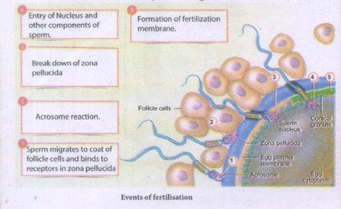
(i) Fertilisation occurs when a haploid sperm fuses with a haploid ovum to form a fertilized egg or diploid zygote.
(ii) The sperms deposited in the female reproductive tract undergo capacitation, which is a biochemical event that enables the sperm to penetrate and fertilise the egg.
(iii) Fertilization occurs only if the ovum and sperms are transported simultaneously to the ampullary isthmic junction of the fallopian tube.
(Iv) Before a sperm can enter the egg, it must penetrate the multiple layers of granulosa (follicular) cells which are around the ovum forming the corona radiata. The follicular cells are held together by an adhesive cementing substance called hyaluronic acid.
(v) The acrosomal membrane disintegrates releasing the proteolytic enzyme, hyaluronidase during sperm entry through the corona radiata and zona pellucida. This is called acrosomal reaction.
(vi) Once fertilisation is accomplished, cortical granules from the cytoplasm of the ovum form a barrier called the fertilisation membrane around the ovum preventing further penetration of other sperms. Thus polyspermy is prevented.
This is followed by cleavage
14.
Structure of human spermatozoan:
(i) The human sperm is a microscopic, flagellated and motile gamete. The whole body of the sperm is enveloped by plasma membrane and is composed of a head, neck and a tail.
(ii) The head comprises of two parts namely acrosome and nucleus.
(iii) Acrosome is a small cap like pointed structure present at the tip of the nucleus and is formed mainly from the golgi body of the spermatid.
(iv) It contains hyaluronidase, a proteolytic enzyme, popularly known as sperm lysin which helps to penetrate the ovum during fertilisation.
(v) The neck is very short and is present between the head and the middle piece. It contains the proximal centriole towards the nucleus which plays a role in the first division of zygote and the distal centriole gives rise to the axial filament of the sperm.
(vi) The middle piece possesses mitochondria spirally twisted around the axial filament called mitochondrial spiral or nebenkern. It produces energy in the form of ATP molecules for the movement of sperms.
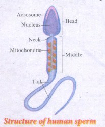
(vii) The tail is the longest part of the sperm and is slender and tapering.
(viii) It is formed of a central axial filament or axoneme and an outer protoplasmic sheath. The lashing movements of the tail push the sperm forward.
15.
| Spermatogenesis | Oogenesis |
| It takes place in the seminiferous tubules of testis |
It occurs in the follicles of the ovary |
| It begins at puberty in males | In females it initiates in the foetal stages. |
| Spermatogonia are the sperm mother cells and many of them undergo maturation phase to form sperms. |
Oogonia is the the egg mother cell and only one cell undergoes maturation phase at a time except in cases of fraternal twins. |
| I & II Mieotic divisions are equal and each spermatogonia forms 4 haploid spermatids. |
I & II meiotic divisions are unequal and finally only one haploid ovum and 3 small haploid polar bodies are formed |
| The spermatids undergo a stage called Spermiogenesis to become functional sperm. |
No such changes are observed. |
| The meiotic divisions are completed and spermaticly are formed. |
The second meiotic division is never completed it fertilization does not take lace. |

12th Standard Syllabus & Materials
12th Standard
TN 12th Computer Applications வலையமைப்பு வடமிடல் Sample Question Papers Study Material - QB365 Set A
NEW12th Standard
TN 12th Computer Applications களப்பெயர் முறைமை (DNS) Sample Question Papers Study Material - QB365 Set A
NEW12th Standard
TN 12th Computer Applications வலையமைப்பு எடுத்துக்காட்டுகள் மற்றும் நெறிமுறைகள் Sample Question Papers Study Material - QB365 Set A
NEW12th Standard
TN 12th Computer Applications கணினி வலையமைப்பு ஓர் அறிமுகம் Sample Question Papers Study Material - QB365 Set A



Tamilnadu Stateboard 12th Standard Subjects

Maths

Chemistry

Physics
Biology

Computer Science

Business Maths and Statistics

Economics

Commerce

Accountancy

History

Computer Applications
Biology
Computer Technology
Computer Applications
Computer Science
Business Maths and Statistics
Commerce
Economics
Maths
Chemistry
Physics
Computer Technology
History
Accountancy

Tamil

English

French
Tamilnadu Stateboard Standards