 100% FREE ACCESS
12th Standard Syllabus & Materials
12th Standard
TN 12th Computer Applications மின்னணு தரவு பரிமாற்றம் Sample Question Papers Study Material - QB365 Set A
NEW12th Standard
TN 12th Computer Applications மின் - வணிக பாதுகாப்பு அமைப்புகள் Sample Question Papers Study Material - QB365 Set A
NEW12th Standard
TN 12th Computer Applications மின்னணு செலுத்தல் முறைகள் Sample Question Papers Study Material - QB365 Set A
NEW12th Standard
TN 12th Computer Applications மின் - வணிகம் Sample Question Papers Study Material - QB365 Set A
NEW12th Standard
TN 12th Computer Applications திறந்த மூல கருத்துருக்கள் Sample Question Papers Study Material - QB365 Set A
NEW12th Standard
TN 12th Computer Applications வலையமைப்பு வடமிடல் Sample Question Papers Study Material - QB365 Set A
Published on: 19/06/2021
QB365 provides detailed and simple solution for every Creative Questions in class 12 Biology Subject. It will helps to get more idea about question pattern in every Creative questions with solution.
Download Tamil Nadu 12th Standard Biology question papers, model tests, one-mark questions, important questions, and public exam papers in PDF format. Free study materials and answer keys for TN State Board students.
Questions + Answers key
Take MCQ Biology Test1.
Write a note on embryonic membranes
2.
Describe the spermatogenesis with diagram.
3.
Describe the structure of human ovary.
4.
Explain the process of fertilization in human beings.
5.
Differentiate Spermatogenesis and oogenesis.
1.
The extra embryonic membranes include amnion, yolk sac, allantois, and chorion. They protect the embryo from desiccation, mechanical shock, absorption of nutrients, gaseous exchange and placental formation.
| Embryonic membrane | Function | |
| a. | Amnion | Innermost two-layered membrane with amniotic fluid providing buoyancy to embryo from injury regulates temperature and acts as a medium for fetal movement. |
| b. | Chorion | Outermost layer covering and protecting the embryo. |
| c. | Allantois | Forms an outpocketing of embryonic tissue at the tail of yolk sac which forms umbilical cord that links embryo to placenta and finally becomes the part of urinary bladder |
| d. | Yolk sac | Forms a part of gut and acts a source of earliest blood cells and blood vessels |
2.
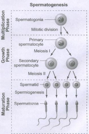
Spermatogenesis is the sequence of events in the seminiferous tubules of the testes that produce the male gametes, the sperms. During development, the primordial germ cells migrate into the testes and become immature germ cells called sperm mother cells or spermatogonia in the inner surfaces of the seminiferous tubules. The spermatogonia begin to undergo mitotic division at puberty and continue throughout life. In the first stage of spermatogenesis, the spermatogonia migrate among Sertoli cells towards the central lumen of the seminiferous tubule and become modified and enlarged to form primary spermatocytes which are diploid with 23 pairs i.e., 46 chromosomes. Some of the primary spermatocytes undergo first meiotic division to form two secondary spermatocytes which are haploid with 23 chromosomes each. The secondary spermatocytes undergo second meiotic division to produce four haploid spermatids. The spermatids are transformed into mature spermatozoa (sperms) by the process called spermiogenesis. Sperms are finally released into the cavity of seminiferous tubules by a process called spermiation. The whole process of spermatogenesis takes about 64 days. At any given time, different regions of the seminiferous tubules contain spermatocytes in different stages of development. The sperm production remains nearly constant at a rate of about 200 million sperms per day. Spermatogenesis starts at the age of puberty and is initiated due to the increase in the release of Gonadotropin-Releasing Hormone (GnRH) by the hypothalamus. GnRH acts on the anterior pituitary gland and stimulates the secretion of two gonadotropins namely Follicle Stimulating Hormone (FSH) and Luteinizing Hormone (LH). FSH stimulates testicular growth and enhances the production of Androgen Binding Protein (ABP) by the Sertoli cells and helps in the process of spermiogenesis. LH acts on the Leydig cells and stimulates the synthesis of testosterone which in turn stimulates the process of spermatogenesis.
3.
(i) Ovaries are the primary female sex organ producing eggs (ovum).
(ii) They are located one on each side of lower abdomen (pelvis).
(iii) The ovary is attached in pelvic wall uterus by an ovarian ligament called mesovarium.
(iv) It is an elliptical structure of 2-4 cm long
(v) Each ovary is covered by thin cuboidal germinal epithelium encloses ovarian stroma.
(vi) Below germinal epithelium is a dense connective tissue called tunica albuginea.
(vii) The stroma is differentiated into an outer cortex and an inner medulla.
(viii) The cortex is dense and granular due to follicular cells at varying development stages.
(ix) The medulla is a loose connective tissue with blood vessels, lymph vessels and nerve fibres.
4.
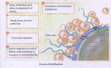
(i) Fertilisation occurs when a haploid sperm fuses with a haploid ovum to form a fertilized egg or diploid zygote.
(ii) The sperms deposited in the female reproductive tract undergo capacitation, which is a biochemical event that enables the sperm to penetrate and fertilise the egg.
(iii) Fertilization occurs only if the ovum and sperms are transported simultaneously to the ampullary isthmic junction of the fallopian tube.
(Iv) Before a sperm can enter the egg, it must penetrate the multiple layers of granulosa (follicular) cells which are around the ovum forming the corona radiata. The follicular cells are held together by an adhesive cementing substance called hyaluronic acid.
(v) The acrosomal membrane disintegrates releasing the proteolytic enzyme, hyaluronidase during sperm entry through the corona radiata and zona pellucida. This is called acrosomal reaction.
(vi) Once fertilisation is accomplished, cortical granules from the cytoplasm of the ovum form a barrier called the fertilisation membrane around the ovum preventing further penetration of other sperms. Thus polyspermy is prevented.
This is followed by cleavage
5.
| Spermatogenesis | Oogenesis |
| It takes place in the seminiferous tubules of testis |
It occurs in the follicles of the ovary |
| It begins at puberty in males | In females it initiates in the foetal stages. |
| Spermatogonia are the sperm mother cells and many of them undergo maturation phase to form sperms. |
Oogonia is the the egg mother cell and only one cell undergoes maturation phase at a time except in cases of fraternal twins. |
| I & II Mieotic divisions are equal and each spermatogonia forms 4 haploid spermatids. |
I & II meiotic divisions are unequal and finally only one haploid ovum and 3 small haploid polar bodies are formed |
| The spermatids undergo a stage called Spermiogenesis to become functional sperm. |
No such changes are observed. |
| The meiotic divisions are completed and spermaticly are formed. |
The second meiotic division is never completed it fertilization does not take lace. |

12th Standard Syllabus & Materials
12th Standard
TN 12th Computer Applications களப்பெயர் முறைமை (DNS) Sample Question Papers Study Material - QB365 Set A
NEW12th Standard
TN 12th Computer Applications வலையமைப்பு எடுத்துக்காட்டுகள் மற்றும் நெறிமுறைகள் Sample Question Papers Study Material - QB365 Set A
NEW12th Standard
TN 12th Computer Applications கணினி வலையமைப்பு ஓர் அறிமுகம் Sample Question Papers Study Material - QB365 Set A
NEW12th Standard
TN 12th Computer Applications PHP-உடன் MySQL-ஐ இணைத்தல் Sample Question Papers Study Material - QB365 Set A



Tamilnadu Stateboard 12th Standard Subjects

Maths

Chemistry

Physics
Biology

Computer Science

Business Maths and Statistics

Economics

Commerce

Accountancy

History

Computer Applications
Biology
Computer Technology
Computer Applications
Computer Science
Business Maths and Statistics
Commerce
Economics
Maths
Chemistry
Physics
Computer Technology
History
Accountancy

Tamil

English

French
Tamilnadu Stateboard Standards