 100% FREE ACCESS
11th Standard CBSE Syllabus & Materials
11th Standard CBSE
CBSE 11th Economics PART-A - Presentation of Data - New Sample Question Papers Study Material - QB365 Set A
NEW11th Standard CBSE
CBSE 11th Economics PART-A - Organisation of Data - New Sample Question Papers Study Material - QB365 Set A
NEW11th Standard CBSE
CBSE 11th Economics PART-A - Collection of Data - New Sample Question Papers Study Material - QB365 Set A
NEW11th Standard CBSE
CBSE 11th Economics PART-A - Introduction to Economics and Statistics - New Sample Question Papers Study Material - QB365 Set A
NEW11th Standard CBSE
CBSE 11th Business Studies International Trade Sample Question Papers Study Material - QB365 Set A
NEW11th Standard CBSE
CBSE 11th Business Studies Evolution and Fundamentals of Business Sample Question Papers Study Material - QB365 Set A

Published on: 21/10/2019
Body Fluids and Circulation
Download CBSE Class 11th Standard CBSE Biology question papers, sample papers, important questions, and previous year solved papers in PDF format. Get free study materials, NCERT solutions, and exam preparation resources for Class 11th Standard CBSE Biology
Questions + Answers key
Take MCQ Biology Test
1.
Describe conduction of heartbeat with the help of suitable diagram.
2.
Define blood. Give detail of various formed elements of blood.
3.
Describe circulatory pathways in animals.
4.
With the help of suitable diagram describe the structure of human heart.
5.
Draw a standard ECG and explain the different segments in it.
6.
Describe the evolutionary change in the pattern of heart among the vertebrates.
7.
Write the differences between :
(a) Blood and Lymph
(b) Open and Closed system of circulation
(c) Systole and Diastole
(d) P-wave and T-wave
8.
Why do we consider blood as a connective tissue?
9.
Explain different types of blood groups and donor compatibility making a table.
1.
Conduction of Heart Beat:
The entire heart is made of cardiac muscles. The walls of ventricles are much thicker than that of the atria. A specialised cardiac musculature called the nodal tissue is also distributed in the heart.
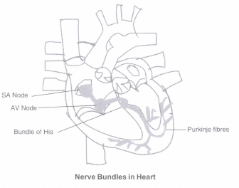
SA Node. A patch of nodal tissue is present in the right upper corner of the right atrium called the sino-atrial node (SAN).
AV Node. Another mass of nodal tissue is seen in the lower left corner of the right atrium close to the atria-ventricular septum called the atria-ventricular node (AVN).
Bundle of His. A bundle of nodal fibres, atrioventricular bundle (AV bundle) continues from the AVN which passes through the atria-ventricular septa to emerge on the top of the interventricular septum and immediately divides into a right and left bundle. These branches give rise to minute fibres throughout the ventricular musculature of the respective sides and are called purkinje fibres. These fibres along with right and left bundles are known as bundle of HIS.
Generation of Heart Beat. The nodal musculature has the ability to generate action potentials without any external stimuli, i.e., it is autoexcitable. However, the number of action potentials that could be generated in a minute vary at different parts of the nodal system. The SA Node can generate the maximum number of action potentials, i.e., 70-75 min-1, and is responsible for initiating and maintaining the rhythmic contractile activity or beating of the heart. Therefore, it is called the pacemaker. Our heart normally beats 70-75 times in a minute (average 72 beats per minute).
2.
Blood is a special connective tissue consisting of a fluid matrix, plasma, and formed elements.
Formed Elements:
Erythrocytes, leucocytes and platelets are collectively called formed elements and they constitute nearly 45 per cent of the blood.
(a) Erythrocytes. They are also known as Red Blood Cells (RBC). They are the most abundant of all the cells in blood. A healthy adult man has, on an average, 5 millions to 5.5 millions of RBCs mm-3 of blood. RBCs are formed in the red bone marrow in the adults. RBCs are devoid of nucleus in most of the mammals and are biconcave in shape. They have a red coloured, iron-containing complex protein called haemoglobin, hence the colour and name of these cells. A healthy individual has 12-16 gms of haemoglobin in every 100 ml of blood. These molecules play a significant role in transport of respiratory gases. RBCs have an average lifespan of 120 days after which they are destroyed in the spleen. Hence, spleen is also known as the graveyard of RBCs.
(b) Leucocytes. They are also known as White Blood Cells (WBC)as they are colourless due to the lack of haemoglobin. They are nucleated and are relatively lesser in number which averages 6000-8000
mm-3 of blood. Leucocytes are generally short lived.
There are two main categories of WBCs:
1. Granulocytes, e.g., neutrophils, eosinophils and basophils
2. Agranulocytes. e.g., Lymphocytes and monocytes.
1. Neutrophils are the most abundant cells (60-65 per cent) of the total WBCs and basophils are the least (0.5-1 per cent) among them. Neutrophils and monocytes (6-8 per cent) are phagocytic cells which destroy foreign organisms entering the body.
2. Basophils secrete histamine, serotonin, heparin, etc., and are involved in inflammatory reactions.
3. Eosinophils (2-3 per cent) resist infections and are also associated with allergic reactions.
4. Lymphocytes (20-25 per cent) are of two major types - 'B' and "T" forms. Both B and T lymphocytes are responsible for immune responses of the body.
3.
The circulatory patterns are of two types - open or closed.
Open Circulatory System. In open circulatory system blood pumped by the heart passes through large vessels into open spaces or body cavities called sinuses. Arthropodas and Molluscs have this type of circulatory system.
Closed Circulatory System. In closed circulatory system blood pumped by the heart is always circulated through a closed network of blood vessels. This pattern is considered to be more advantageous as the flow of fluid can be more precisely regulated. Annelids and chordates have this type of system.
Single Circulation. In fishes, the heart pumps out deoxygenated blood which is oxygenated by the gills and supplied to the body parts from where deoxygenated blood is returned to the heart.
Incomplete Double Circulation. In amphibians and reptiles, the left atrium receives oxygenated blood from the gills/lungs/ skin and the right atrium gets the deoxygenated blood from other body parts. However, they get mixed up in the single ventricle which pumps out mixed blood.
Complete Double Circulation. In birds and mammals, oxygenated and deoxygenated blood received by the left and right atria respectively passes on to the ventricles of the same sides. The ventricles pump it out without any mixing up, i.e., two separate circulatory pathways are present in these organisms, hence, these animals have double circulation.
4.
Heart
Location and Size. Heart, the mesodermally derived organ, is situated in the thoracic cavity, in between the two lungs, slightly tilted to the left. It has the size of a clenched fist.
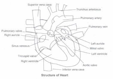
Structure. The heart is protected by a double-walled membranous bag, pericardium, enclosing the pericardial fluid. Heart has four chambers:
(a) Two relatively small upper chambers called atria and
(b) two larger lower chambers called ventricles.
Septum. A thin, muscular wall called the interatrial septum separates the right and the left atria, whereas a thick-walled, the inter-ventricular septum, separates the left and the right ventricles The atrium and the ventricle of the same side are also separated by a thick fibrous tissue called the atrio-ventricular septum. However, each of these septa are provided with an opening through which the two chambers of the same side are connected.
Valves. The opening between the right atrium and the right ventricle is guarded by a valve formed of three muscular flaps or cusps, the tricuspid valve, whereas a bicuspid or mitral valve guards the opening between the left atrium and the left ventricle. The openings of the right and the left ventricles into the pulmonary artery and the aorta respectively are provided with the semilunar valves. The valves in the heart allows the flow of blood only in one direction, i.e., from the atria to the ventricles and from the ventricles to the pulmonary artery or aorta. These valves prevent any backward flow.
5.
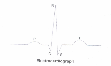
The P-wave represents the electrical excitation (or depolarisation) of the atria, which leads to the contraction of both the atria.
The QRS complex represents the depolarisation of the ventricles, which initiates the ventricular contraction. The contraction starts shortly after Q and marks the beginning of the systole.
The T-wave represents the return of the .ventricles from excited to normal state (repolarisation). The end of the T-wave marks the end of systole. Obviously, by counting the number of QRS complexes that occur in a given time period, one can determine the heartbeat rate of an individual. Since the ECGs obtained from different individuals have roughly the same shape for a given lead configuration, any deviation from this shape indicates a possible abnormality or disease. Hence, it is of a great clinical significance.
6.
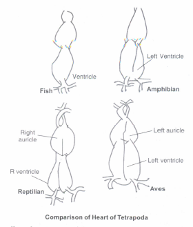
As is clear from the following diagram the heart of fish has two chambers. This means there is no separate circulation for oxygenated and deoxygenated blood. There is separation of two chambers in the atrium of amphibians. This has further evolved to partial separation of ventricle as well in reptiles. Finally, in birds, there is complete separation of oxygenated and deoxygenated blood circulation with advent of four chambers in the heart. Mammal heart is the most developed having the most efficient double circulatory system.
7.
(a) Blood contains RBCs and hence can transport gases. Lymph doesn't contain RBCs and cannot transport gases. Lymph mainly contains WBCs and play a role in the immune system of the body.
(b) The Open Circulatory System is a system in which fluid (called hemolymph) in a cavity called the hemocoel bathes the organs directly with oxygen and nutrients and there is no distinction between blood and interstitial fluid; this combined fluid is called hemolymph or haemolymph. The cardiovascular systems of humans are closed, meaning that the blood never leaves the network of blood vessels.
(c) Systole is the contraction of heart muscle and diastole is the dilatation of the heart muscle.
(d) Each peak in the ECG is identified with a letter from P to T that corresponds to a specific electrical activity of the heart. The P-wave represents the electrical excitation (or depolarisation) of the atria, which leads to the contraction of both the atria. The QRS complex represents the depolarisation of the ventricles, which initiates the ventricular contraction. The contraction starts shortly after Q and marks the beginning of the systole. The T-wave represents the return of the ventricles from excited to normal state (repolarisation). The end of the T-wave marks the end of systole.
8.
Connective Tissue: It is largely a category of exclusion rather than one with a precise definition, but all or most tissues in this category are similarly:
(a) Involved in structure and support.
(b) Derived from mesoderm, usually.
(c) Characterized largely by the traits of non-living tissue.
Blood is considered a connective tissue for two basic reasons:
(i) embryologically, it has the same origin (mesodermal) as do the other connective tissue types and
(ii) blood connects the body systems together bringing the needed oxygen, nutrients, hormones and other signaling molecules, and removing the wastes. As the name implies, connective tissue serves a "connecting" function. It supports and binds other tissues. Unlike epithelial tissue, connective tissue typically has cells scattered throughout an extracellular matrix. Interestingly enough, blood is considered to be a type of connective tissue. Even though it has a different function in comparison to other connective tissues it does have an extracellular matrix. The matrix is the plasma and erythrocytes, leucocytes and platelets are suspended in the plasma.
9.
Two groupings, i.e., the ABO and Rh- are widely used all over the world. ABO grouping is based on the presence or absence of two surface antigens (chemicals that can induce immune response) on the RBCs, i.e., A and B. Similarly, the plasma of different individuals contain two natural antibodies (proteins produced in response to antigens).
Blood Groups and Donor Compatibility
| Blood Group | Antigen on RCBs | Antibody in Plasma | Donor's Group |
| A | A | Anti-B | A, O |
| B | B | Anti-A | B, O |
| AB | A, B | Nil | AB, A, B, O |
| O | Nil | Anti- A, B | O |
From the above-mentioned table, it is evident that group 'O' blood can be donated to persons with any other blood group and hence 'O' group individuals are called 'universal donors'. Persons with 'AB' group can accept blood from persons with AB as well as the other groups of blood. Therefore, such persons are called 'universal recipients'.

11th Standard CBSE Syllabus & Materials
11th Standard CBSE
CBSE 11th Business Studies Forms of Business Organisation Sample Question Papers Study Material - QB365 Set A
NEW11th Standard CBSE
CBSE 11th Business Studies Business, Trade and Commerce Sample Question Papers Study Material - QB365 Set A
NEW11th Standard CBSE
CBSE 11th Physics Waves Sample Question Papers Study Material - QB365 Set A
NEW11th Standard CBSE
CBSE 11th Physics Kinetic Theory Sample Question Papers Study Material - QB365 Set A



CBSE 11th Standard CBSE Subjects
CBSE Standards