 100% FREE ACCESS
11th Standard CBSE Syllabus & Materials
11th Standard CBSE
CBSE 11th Economics PART-A - Presentation of Data - New Sample Question Papers Study Material - QB365 Set A
NEW11th Standard CBSE
CBSE 11th Economics PART-A - Organisation of Data - New Sample Question Papers Study Material - QB365 Set A
NEW11th Standard CBSE
CBSE 11th Economics PART-A - Collection of Data - New Sample Question Papers Study Material - QB365 Set A
NEW11th Standard CBSE
CBSE 11th Economics PART-A - Introduction to Economics and Statistics - New Sample Question Papers Study Material - QB365 Set A
NEW11th Standard CBSE
CBSE 11th Business Studies International Trade Sample Question Papers Study Material - QB365 Set A
NEW11th Standard CBSE
CBSE 11th Business Studies Evolution and Fundamentals of Business Sample Question Papers Study Material - QB365 Set A

Published on: 03/10/2019
Structural Organisation in Animals
Download CBSE Class 11th Standard CBSE Biology question papers, sample papers, important questions, and previous year solved papers in PDF format. Get free study materials, NCERT solutions, and exam preparation resources for Class 11th Standard CBSE Biology
Questions + Answers key
Take MCQ Biology Test
1.
Explain the reproductive system in cockroach with the help of suitable diagrams.
2.
Explain morphology of cockroach.
3.
Explain the morphology of an earthworm.
4.
With the help of appropriate diagrams explain various kinds of muscular tissues with their junctions.
5.
Describe various types of epithelial tissues with the help of labelled diagrams.
6.
Distinguish between a male cockroach and female cockroach.
7.
Differences Between Blatta orientalis (oriental cockroach) and peripalnata americana (Aamerican cockroach).
8.
Stratified epithelial cells have limited role in secreation . Justify their role in our skin
1.
Reproductive System. Cockroaches are dioecious and both sexes have well developed reproductive organs.
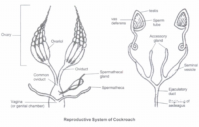
Male Reproductive System:
(i) Male reproductive system consists of a pair of testes lying one on each lateral side in the 4th-6th abdominal segments.
(ii) From each testis arises a thin vas deferens, which opens into ejaculatory duct through seminal vesicle. The ejaculatory duct opens into male gonopore situated ventral to anus.
(iii) A characteristic mushroom shaped gland is present in the 6th-7th abdominal segments which functions as an accessory reproductive gland.
(iv) The external genitalia are represented by male gonapophysis or phallomere (chitinous asymmetrical structures, surrounding the male gonopore).
(v) The sperms are stored in the seminal vesicles and are glued together in the form of bundles called spermatophores which are discharged during copulation.
Female Reproductive System:
(i) The female reproductive system consists of two large ovaries, lying laterally in the 2nd - 6th abdominal segments. Each ovary is formed of a group of eight ovarian tubules or ovarioles, containing a chain of developing ova.
(ii) Oviducts of each ovary unite into a single median oviduct (also called vagina) which opens into the genital chamber.
(iii) A pair of spermatheca is present in the 6th segment which opens into the genital chamber.
Fertilization:
(i) Sperms are transferred through spermatophores.
(ii) Their fertilised eggs are encased in capsules called oothecae. Ootheca is a dark reddish to blackish brown capsule, about 3/8" (8 mm) long. They are dropped or glued to a suitable surface, usually in a crack or crevice of high relative humidity near a food source.
(iii) On an average, females produce 9-10 oothecae, each containing 14-16 eggs.
(iv) The development of P. Americana is paurometabolous, meaning there is development through nymphal stage. The nymphs look very much like adults. The nymph grows by moulting about 13 times to reach the adult form. The next to last nymphal stage has wing pads but only adult cockroaches have wings.
2.
Morphology of Cockroach
The adults of the common species of cockroach, Periplaneta Americana are about 34-53 mm long with wings that extend beyond the tip of the abdomen in males. The body of the cockroach is segmented and divisible into three distinct regions
(i) head,
(ii) thorax and
(iii) abdomen.
Exoskeleton. The entire body is covered by a hard chitinous exoskeleton (brown in colour). In each segment, exoskeleton has hardened plates called sclerites (tergites dorsally and sternites ventrally) that are joined to each other by a thin and flexible articular membrane (arthrodial membrane).
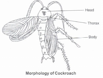
Head:
(i) Head is triangular in shape and lies anteriorly at right angles to the longitudinal body axis.
(ii) It is formed by the fusion of six segments and shows great mobility in all directions due to flexible neck.
(iii) The head capsule bears a pair of compound eyes.
(iv) A pair of thread like antennae arises from membranous sockets lying in front of eyes. Antennae have sensory receptors that help in monitoring the environment.
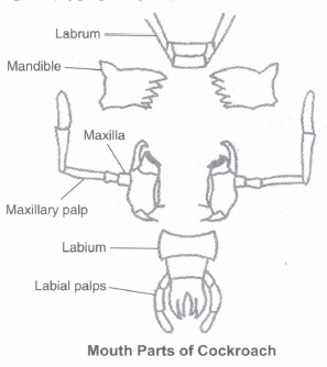
Mouth Parts. Anterior end of the head bears appendages forming biting and chewing type of mouth parts. The mouthparts consisting of a labrum (upper lip), a pair of mandibles, a pair of maxillae and a labium (lower lip). A median flexible lobe, acting as tongue (hypopharynx), lies within the cavity enclosed by the mouthparts.
Thorax. Thorax consists of three parts
(i) prothorax,
(ii) mesothorax and
(iii) metathorax.
The head is connected with thorax by a short extension of the prothorax known as the neck. Each thoracic segment bears a pair of walking legs. The first pair of wings arises from mesothorax and the second pair from metathorax. Forewings (mesothoracic) called tegmina are opaque dark and leathery and cover the hind wings when at rest. The hind wings are transparent, membranous and are used in flight.
Abdomen:
(i) The abdomen in both males and females consists of 10 segments.
(ii) In females, the 7th sternum is boat shaped and together with the 8th and 9th sterna forms a brood or genital pouch whose anterior part contains female gonopore, spermathecal pores and collateral glands.
(iii) In males, genital pouch or chamber lies at the hind end of abdomen bounded dorsally by 9th and 10th terga and ventrally by the 9th sternum. It contains dorsal anus, ventral male genital pore and gonapophysis. Males bear a pair of short, threadlike anal styles which are absent in females.
(iv) In both sexes, the 10th segment bears a pair of jointed filamentous structures called anal cerci.
3.
Morphology of Earthworm:
Body. Earthworms have long cylindrical body. The body is divided into more than hundred short segments which are similar (metameres about 100-120 in number).
Dorsal Surface. The dorsal surface of the body is marked by a dark median mid dorsal line (dorsal blood vessel) along the longitudinal axis of the body.
Ventral Surface. The ventral surface is distinguished by th~ presence of genital openings (pores).
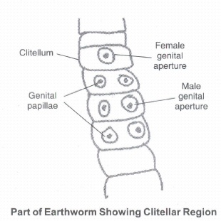
Segments. In a mature worm, segments 14-16 are covered by a prominent dark band of glandular tissue called clitellum. Thus the body is divisible into three prominent regions
(i) preclitellar
(ii) clitellar and
(iii) postclitellar segments.
Buccal Region. Anterior end consists of the mouth and the prostomium, a lobe which serves as a covering for the mouth and as a wedge to force open cracks in the soil into which the earthworm may crawl. The prostomium is sensory in function. The first body segment is called the peristomium (buccal segment) which contains the mouth.
Genital Openings. Four pairs of spermathecal apertures are situated on the ventra-lateral sides of the intersegmental grooves, i.e., 5th-9th segments. A single female genital pore is present in the mid-ventral line of 14th segment. A pair of male genital pores are present on the ventra-lateral sides of the 18th segment
Locomotion. In each body segment, except the first, last and clitellum, there are rows of S-shaped setae, embedded in the epidermal pits in the middle of each segment. Setae can be extended or retracted. Their principal role is in locomotion.
4.
Muscle Tissue
(i) Each muscle is made of many long, cylindrical fibres arranged in parallel arrays.
(ii) These fibres are composed of numerous fine fibrils, called myofibrils.
(iii) Muscle fibres contract (shorten) in response to stimulation, then relax (lengthen) and return to their uncontracted state in a coordinated fashion.
(iv)Their action moves the body to adjust to the changes in the environment and to maintain the positions of the various parts of the body.
Muscles are of three types:
(i) Skeletal,
(ii) Smooth, and
(iii) Cardiac.
Skeletal or Striated Muscle. Skeletal muscle tissue is closely attached to skeletal bones. Striated or skeletal muscle fibres are bundled together in a parallel fashion. A sheath of tough connective tissue encloses several bundles of muscle fibres.
Function. Skeletal muscles are 'voluntary' as their function is under the conscious control. They facilitate articulation of limbs, neck, torso, etc.
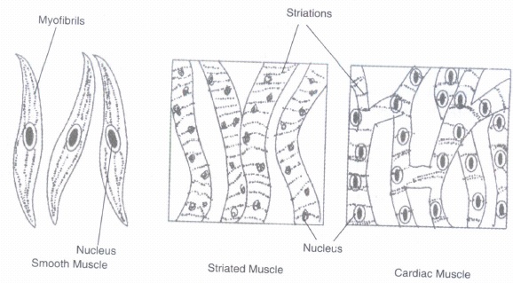
Smooth Muscle. The smooth muscle fibres taper at both ends (fusiform) and do not show striations. Cell junctions hold them together and they are bundled together in a connective tissue sheath. The wall of internal organs such as the blood vessels, stomach and intestine contains this type of muscle tissue.
Function. Smooth muscles are 'involuntary' as their functioning cannot be consciously controlled.
Cardiac Muscle. Cardiac muscle tissue is a contractile tissue present only in the heart. Cell junctions fuse the plasma membranes of cardiac muscle cells and make them stick together. Communication junctions (intercalated discs) at some fusion points allow the cells to contract as a unit, i.e., when one cell receives a signal to contract, its neighbours are also stimulated to contract.
Function. Facilitate pumping action of heart.
5.
Epithelial Tissue
Epithelial tissues provide covering to the inner and outer lining of various organs. The cells of epithelial tissues are compactly packed with little intercellular matrix. There are two types of epithelial tissues:
(a) Simple Epithelium. Simple epithelium is composed of a single layer of cells and functions as a lining for body cavities, ducts, and tubes.
(b) Compound Epithelium. The compound epithelium consists of two or more cell layers and has protective function as it does in our skin. They cover the dry surface of the skin, the moist surface of buccal cavity, pharynx, inner lining of ducts of salivary glands and of pancreatic ducts.
On the basis of structural modification of the cells, simple epithelium is further divided into three types. These are:
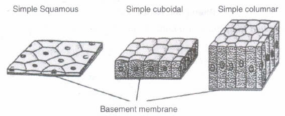
1. Squamous. The squamous epithelium is made of a single thin layer of flattened cells with irregular boundaries. They are found in the walls of blood vessels and air sacs of lungs and are involved in functions, like forming a diffusion boundary.
2. Cuboidal. The cuboidal epithelium is composed of a single layer of cubelike cells. This is commonly found in ducts of glands and tubular parts of nephrons in kidneys and its main functions are secretion and absorption. The epithelium of proximal convoluted tubule (PCT) of nephron in the kidney has microvilli.
3. Columnar. The columnar epithelium is composed of a single layer of tall and slender cells. Their nuclei are located at the base. Free surface may have microvilli. They are found in the lining of stomach and intestine and help in secretion and absorption. If the columnar or cuboidal cells bear cilia on their free surface they are called ciliated epithelium. Their function is to move particles or mucus in a specific direction over the epithelium. They are mainly present in the inner surface of hollow organs like bronchioles and fallopian tubes.
6.
Differences between male and female cockroach are
| Male Cockroach | Female Cockroach |
| Abdomen is long and narrow | Abdomen is short and broad |
| All nine sterna are visible | Only seven sterna are visible |
| Anal styles are present | Anal styles are absent |
| 7 th tergum coves 8th tergum | 7th tergum covers 8th and 9th terga. |
| Brood pouch is absent | B |
| Antennae are longer in size | Antennae are smaller in size |
| Wings extend beyond the tip of abdomen | Wings extend upto the end of abdomen |
7.
Differences between Blatta orientals and Periplaneta americana are
| Blatta orientals | Periplanata Americana |
| Brownish-black to black coloured | Shining brown-cloured |
| 20-25 mm, smaller in size | 35-40 mm, larger in size |
| Less capable of lying | Good capacity of flying |
| Forewings are vestigial Hindwings are absent. | In both sexes, both pairs of wings are well-developed. |
| Ootheca is black-coloured | Oootheca is grey-coloured |
8.
Features: Stratified epithelium is made up of many layers of cells. In the skin the innermost layers are made of cuboidal or squamous cells. The outer layers are made of keratinised epithelium. Keratinised epithelium cells have lost their nucleus and contain a lot of keratin that help in protection.
Role in the skin:
Keratinised stratified epithelium is found in the skin because-
1. This epithelium contains dead cells with a lot of insoluble protein called keratin therefore providing the epidermis of the skin with protection against germs and desiccation.
2. They have a minimum role in secretions in the skin.
3. The secretions in the skin are due to glandular epithelium and not by the keratinised stratified epithelium.


11th Standard CBSE Syllabus & Materials
11th Standard CBSE
CBSE 11th Business Studies Forms of Business Organisation Sample Question Papers Study Material - QB365 Set A
NEW11th Standard CBSE
CBSE 11th Business Studies Business, Trade and Commerce Sample Question Papers Study Material - QB365 Set A
NEW11th Standard CBSE
CBSE 11th Physics Waves Sample Question Papers Study Material - QB365 Set A
NEW11th Standard CBSE
CBSE 11th Physics Kinetic Theory Sample Question Papers Study Material - QB365 Set A



CBSE 11th Standard CBSE Subjects
CBSE Standards