 100% FREE ACCESS
11th Standard CBSE Syllabus & Materials
11th Standard CBSE
CBSE 11th Economics PART-A - Presentation of Data - New Sample Question Papers Study Material - QB365 Set A
NEW11th Standard CBSE
CBSE 11th Economics PART-A - Organisation of Data - New Sample Question Papers Study Material - QB365 Set A
NEW11th Standard CBSE
CBSE 11th Economics PART-A - Collection of Data - New Sample Question Papers Study Material - QB365 Set A
NEW11th Standard CBSE
CBSE 11th Economics PART-A - Introduction to Economics and Statistics - New Sample Question Papers Study Material - QB365 Set A
NEW11th Standard CBSE
CBSE 11th Business Studies International Trade Sample Question Papers Study Material - QB365 Set A
NEW11th Standard CBSE
CBSE 11th Business Studies Evolution and Fundamentals of Business Sample Question Papers Study Material - QB365 Set A

Published on: 21/10/2019
Neural Control and Coordination
Download CBSE Class 11th Standard CBSE Biology question papers, sample papers, important questions, and previous year solved papers in PDF format. Get free study materials, NCERT solutions, and exam preparation resources for Class 11th Standard CBSE Biology
Questions + Answers key
Take MCQ Biology Test
1.
Describe the detail structure of the human ear.
2.
Describe the structure of the human eye with a well labelled diagram.
3.
Briefly describe the structure of the Brain.
4.
Distinguish between:
(a) afferent neurons and efferent neurons
(b) impulse conduction in a myelinated nerve fibre and unmyelinated nerve fibre
(f) cranial nerves and spinal nerves.
5.
Explain the following:
(al Role of Na+ in the generation of action potential.
(b) Mechanism of generation of light-induced impulse in the retina.
(c) Mechanism through which a sound produces a nerve impulse in the inner ear.
6.
Write short notes on the following:
(a) Neural coordination
(b) Forebrain
(c) Midbrain
(d) Hindbrain
(e) Synapse
7.
Draw labelled diagrams of the following:
(a) Neuron
(b) Brain
8.
Explain the following processes:
(a) Polarisation of the membrane of a nerve fibre
(b) Depolarisation of the membrane of a nerve fibre
(c) Transmission of a nerve impulse across a chemical synapse
9.
Compare the following:
(a) Central neural system (CNS) and Peripheral neural system (PNS)
(b) Resting potential and action potential
10.
Briefly describe the structure of the following:
(a) Brain (b) Eye (c) Ear
1.
Structure of Ear
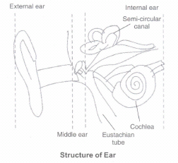
Anatomically, the ear can be divided into three major sections called the outer ear, the middle ear and the inner ear.
Outer Ear. The outer ear consists of the pinna and external auditory meatus (canal). The pinna collects the vibrations in the air which produce sound. The external auditory meatus leads inwards and extends up to the tympanic membrane (the ear drum). There are very fine hairs and wax-secreting sebaceous glands in the skin of the pinna and the meatus. The tympanic membrane is composed of connective tissues covered with skin outside and with mucus membrane inside. Middle Ear. The middle ear contains three ossicles called malleus, incus and stapes which are attached to one another in a chain-like fashion.
The malleus is attached to the tympanic membrane and the stapes is attached to the oval window of the cochlea. The ear ossicles increase the efficiency of transmission of sound waves to the inner ear. A Eustachian tube connects the middle ear cavity with the pharynx. The Eustachian tube helps in equalising the pressures on either side of the ear drum.
Inner Ear.
The fluid-filled inner ear called labyrinth consists of two parts, the bony and the membranous labyrinths. The bony labyrinth is a series of channels. Inside these channels lies the membranous labyrinth, which is surrounded by a fluid called perilymph. The membranous labyrinth is filled with a fluid called endolymph. The coiled portion of the labyrinth is called cochlea.
The membranes constituting cochlea, the reissner's and basilar, divide the surounding perilymph filled bony labyrinth into an upper scala vestibuli and a lower scala tympani. The space within cochlea called scala media is filled with endolymph. At the base of the cochlea, the scala vestibuli ends at the oval window, while the scala tympani terminates at the round window which opens to the middle ear.
The organ of corti is a structure located on the basilar membrane which contains hair cells that act as auditory receptors. The hair cells are present in rows on the internal side of the organ of corti. The basal end of the hair cell is in close contact with the afferent nerve fibres. A large number of processes called stereo cilia are projected from the apical part of each hair cell. Above the rows of the hair cells is a thin elastic membrane called tectorial membrane.
The inner ear also contains a complex system called vestibular apparatus, located above the cochlea. The vestibular apparatus is composed of three semi-circular canals and the otolith organ consisting of the saccule and utricle. Each semicircular canal lies in a different plane at right angles to each other. The membranous canals are suspended in the perilymph of the bony canals. The base of canals is swollen and is called ampulla, which contains a projecting ridge called crista ampullaris which has hair cells. The saccule and utricle contain a projecting ridge called macula. The crista and macula are the specific receptors of the vestibular apparatus responsible for maintenance of balance of the body and posture.
2.
Structure of Eye The adult human eye ball is nearly a spherical structure. The wall of the eye ball is composed of three layers. The external layer is composed of a dense connective tissue and is called the sclera. The anterior portion of this layer is called the cornea. The middle layer, choroid, contains many blood vessels and looks bluish in colour. The choroid layer is thin over the posterior two-thirds of the eye ball, but it becomes thick in the anterior part to form the ciliary body. The ciliary body itself continues forward to form a pigmented and opaque structure called the iris which is the visible coloured portion of the eye. The eye ball contains a transparent crystalline lens which is held in place by ligaments attached to the ciliary body. In front of the lens, the aperture surrounded by the iris is called the pupil. The diameter of the pupil is regulated by the muscle fibres of iris.
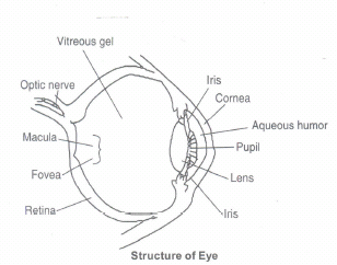
The inner layer is the retina and it contains three layers of cells - from inside to outside - ganglion cells, bipolar cells and photoreceptor cells. There are two types of photoreceptor cells, namely, rods and cones. These cells contain the light-sensitive proteins called the photopigments. The daylight (photopic) vision and colour vision are functions of cones and the twilight (scotopic) vision is the function of the rods. The rods contain a purplish-red protein called the rhodopsin or visual purple, which contains a derivative of Vitamin A. In the human eye, there are three types of cones which possess their own characteristic photopigments that respond to red, green and blue lights. The sensations of different colours are produced by various combinations of these cones and their photopigments. When these cones are stimulated equally, a sensation of white light is produced. The optic nerves leave the eye and the retinal blood vessels enter it at a point medial to and slightly above the posterior pole of the eye ball. Photoreceptor cells are not present in that region and hence it is called the blind spot. At the posterior pole of the eye lateral to the blind spot, here is a yellowish pigmented spot called macula lutea with a central pit called the fovea. The fovea is a thinned-out portion of the retina where only the cones are densely packed. It is the point where the visual acuity (resolution) is the greatest. The space between the cornea and the lens is called the aqueous chamber and contains a thin watery fluid called aqueous humor. The pace between the lens and the retina is called the vitreous chamber and is filled with a transparent gel called vitreous humor.
3.
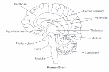
The structure of Brain :
The human brain is divisible into three main parts
1) Forebrain :
It consist of cerebrum, thalamus and hypothalamus.
i) Cerebrum It is the largest and most complex of all the parts of the human brain. A deep cleft divides the cerebrum longitudinally into two equal halves. These are termed as the left and right cerebral hemispheres. These hemispheres are connected by a large band of myelinated fibres, the corpus callosum.
The outer cover of cerebral hemisphere is called cerebral cortex. The cerebral cortex is referred to as grey matter due to its greyish appearance (as neuron cell bodies are concentrated here). The cerebral cortex is greatly folded.
Beneath the grey matter, there are millions of myelinated nerve fibers, which constitute the inner part of the cerebral hemisphere.
The large concentration of myelinated nerve fibres gives this tissue an opaque white appearance. Hence, it is called white matter.
In each cerebral hemisphere, there are three types of functional areas
Sensory Areas : It receives impulses from the receptors.
Motor Areas : Transmit impulses to the effectors.
Association Areas : These are large regions that are neither clearly sensory junction nor motor in They interpret the input, store the input and initiate a response in light of similar past experience. Thus, these areas are responsible for complex functions like memory, communication and other intersensory associations.
(ii) Thalamus : The cerebrum wraps around a structure called thalamus, which is a major coordinating centre for sensory and motor signalling.
(iii) Hypothalamus : It lies at the base of thalamus contains a number of centres, which control body temperature, urge for eating and drinking. It also contains several groups of neurosecretory cells, which secrete hormones called hypothalamic hormones.
The inner parts of cerebral hemispheres and a group of deeply associated structures like amygdala, hippocampus, etc. They form complex structure (limbic lobe or limbic system) that are involved in the regulation of sexual behaviour, expression of emotional reactions, c.g. excitement, pleasure, rage and fear and motivation.
2) Midbrain :
The midbrain is located between the thalamus, hypothalamus of the forebrain and pons of the hindbrain. A canal called the cerebral aqueduct passes through the midbrain.
The dorsal portion of the midbrain mainly consists of two pairs (i.e., four) of rounded swellings (lobes) called corpora quadrigemina.
3. Hindbrain :
It consists of pons, cerebellum and medulla.
(i) Pons It consists of fibre tracts that interconnect different regions of the brain.
(ii) Cerebellum It is the second largest part of the human brain (first being the cerebrum). It has very convoluted surface in order to provide the additionalspace for many more neurons.
(iii) Medulla It is also called medulla oblongata. It is connected to the spinal cord and contains centres, which control respiratory rhythm, cardiovascular reflexes, gastric secretions and the postural gestures of the body (vertigo).
4.
(a) The afferent nerve fibres transmit impulses from tissues/organs to the CNS and the efferent fibres transmit regulatory impulses from the CNS to the concerned peripheral tissues/organs.
(b) The evolutionary need for the fast and efficient transduction of electrical signals in neural system resulted in appearance of myelin sheaths around neuronal axons. Myelin sheath reduces membrane capacitance and increases membrane resistance in the inter-node intervals, thus allowing a fast, saltatory movement of action potentials from node to node. Myelination is found mainly in vertebrates, but an analogous system has been discovered in a few invertebrates, such as some species of shrimp. Not all neurons in vertebrates are myelinated; for example, axons of the neurons comprising autonomous (vegetative) neural system are not myelinated in general. The conduction velocity v of myelinated neurons varies roughly linearly with axon diameter whereas the speed of unmyelinated neurons varies roughly as the square root of diameter. Myelin has two important advantages. fast \ conduction speed and energy efficiency. Also, since the ionic currents are confined to the nodes of Ranvier, there is far fewer ions "leak" across the membrane, saving metabolic energy. This saving is a significant selective advantage, since the human neural system uses approximately 20% of the body's metabolic energy.
(f) Cranial nerves are nerves that emerge directly from the brain stem in contrast to spinal nerves which emerge from segments of the spinal cord. Peripheral nerves are separated to achieve segmental innervation, cranial nerves are divided to serve one or a few specific functions in wider anatomical territories.
5.
(a) The sodium-potassium pump sends 3Na+ outside for every 2K+ inside the axon. This helps in maintaining a higher concentration of Na+ outside the axonal membrane. This further helps maintain the potential difference across the membrane, which is necessary for initiation of action potential.
(b) Light induces dissociation of the retinal from opsin resulting in changes in the structure of the opsin. This causes membrane permeability changes. As a result, potential differences are generated in the photoreceptor cells. This produces a signal that generates action potentials in the ganglion cells through the bipolar cells.
(c) The hollow channels of the inner ear are filled with liquid, and contain a sensory epithelium that is studded with hair cells. The microscopic "hairs" of these cells are structural protein filaments that project out into the fluid. The hair cells are mechanoreceptors that release a chemical neurotransmitter when stimulated. Sound waves moving through fluid push the filaments; if the filaments bend over enough it causes the hair cells to fire. In this way, sound waves are transformed into nerve impulses.
6.
(a) Neural Coordination. The functions of the organs/organ systems in our body must be coordinated to maintain homeostasis. Coordination is the process through which two or more organs interact and complement the functions of one another. For example, when we do physical exercises, the energy demand is increased for maintaining an increased muscular activity. The supply of oxygen is also increased. The increased supply of oxygen necessitates an increase in the rate of respiration, heartbeat and increased blood flow via blood vessels. When physical exercise is stopped, the activities of nerves, lungs, heart and kidney gradually return to their normal conditions. Thus, the functions of muscles, lungs, heart, blood vessels, kidney and other organs are coordinated while performing physical exercises. In our body, the neural system and the endocrine system jointly coordinate and integrate all the activities of the organs so that they function in a synchronised fashion.
(b) Forebrain. The forebrain consists of cerebrum, thalamus and hypothalamus. Cerebrum forms the major part of the human brain. A deep cleft divides the cerebrum longitudinally into two halves, which are termed as the left and right cerebral hemispheres. The hemispheres are connected by a tract of nerve fibres called corpus callosum. The cerebral cortex contains motor areas, sensory areas and large regions that are neither clearly sensory nor motor in function. These regions called as the association areas are responsible for complex functions like intersensory associations, memory and communication. The cerebrum wraps around a structure called thalamus, which is a major coordinating centre for sensory and motor signaling. Another very important part of the brain called hypothalamus lies at the base of the thalamus. The hypothalamus contains a number of centres which control body temperature, urge for eating and drinking. It also contains several groups of neurosecretory cells, which secrete hormones called hypothalamic hormones. The inner parts of cerebral hemispheres and a group of associated deep structures like amygdala, hippocampus, etc., form a complex structure called the limbic lobe or limbic system. Along with the hypothalamus, it is involved in the regulation of sexual behaviour, expression of emotional reactions (e.g., excitement, pleasure, rage and fear), and motivation.
(c) Midbrain. The midbrain is located between the thalamus/hypothalamus of the forebrain and pons of the hindbrain. Acanal called the cerebral aqueduct passes through the midbrain. The dorsal portion of the midbrain consists mainly of four round swellings (lobes) called corpora quadrigemina. Midbrain and hindbrain form the brain stem.
(d) Hindbrain. The hindbrain comprises pons, cerebellum and medulla (also called the medulla oblongata). Pons consists of fibre tracts that interconnect different regions of the brain. Cerebellum has very convoluted surface in order to provide the additional space for many more neurons. The medulla of the brain is connected to the spinal cord. The medulla contains centres which control respiration, cardiovascular reflexes and gastric secretions.
(e) Synapse. A nerve impulse is transmitted from one neuron to another through junctions called synapses. A synapse is formed by the membranes of a presynaptic neuron and a post-synaptic neuron, which may or may not be separated by a gap called synaptic cleft. There are two types of synapses, namely, electrical synapses and chemical synapses. Electrical Synapse. At electrical synapses, the membranes of pre- and postsynaptic neurons are in very close proximity. Electrical current can flow directly from one neuron into the other across these synapses. Transmission of an impulse across electrical synapses is very similar to impulse conduction along a single axon. Impulse transmission across an electrical synapse is always faster than that across a chemical synapse. Electrical synapses are rare in our system.
7.
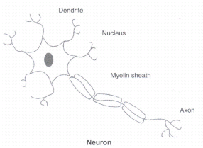
(a) Neuron
(b) Brain
8.
a) Polarisation of the membrane of a nerve fibre. The fluid inside the membrane contains high concentration of K+and negatively charged proteins and low concentration of Na+.
In contrast, the fluid outside the axon contains a low concentration of K+, a high concentration of Na+ and thus forms a concentration gradient. These ionic gradients across the resting membrane are maintained by the active transport of ions by the sodium-potassium pump which transports 3 Na+ outwards for 2 K+ into the cell. As a result, the outer surface of the axonal membrane possesses a positive charge while its inner surface becomes negatively charged and therefore is polarised.
b) Depolarisation of the membrane of a nerve fibre. When a stimulus is applied at a site on the polarised membrane, the membrane at the site A becomes freely permeable to Na+, This leads to a rapid influx of Na+ followed by the reversal of the polarity at that site, i.e., the outer surface of the membrane becomes negatively charged and the inner side becomes positively charged. The polarity of the membrane at the site is thus reversed and hence depolarised.
c) Transmission of a nerve impulse across chemical synapse. At a chemical synapse, the membranes of the pre- and post-synaptic neurons are separated by a fluid-filled space called synaptic cleft. Chemicals called neurotransmitters are involved in the transmission of impulses at these synapses. The axon terminals contain vesicles filled with these neurotransmitters. When an impulse (action potential) arrives at the axon terminal, it stimulates the movement of the synaptic vesicles towards the membrane where they fuse with the plasma membrane and release their neurotransmitters in the synaptic cleft. The released neurotransmitters bind to their specific receptors, present on the post-synaptic membrane. This binding opens ion channels allowing the entry of ions which can generate a new potential in the postsynaptic neuron. The new potential developed may be either excitatory or inhibitory.
9.
(a) Central neural system (CNS) and Peripheral neural system (PNS)
Central neural system (CNS) :
It is the main coordinating centre of the body.
It lies inside the skull.
This includes brain and spinal cord.
Peripheral neural system (PNS) :
It is not the main coordinating centre of the body.body.
It is present outside the skull in the peripheral areas of the body.
This includes all the nerves of the bodyassociated with the CNS (brain and spinalcord).
(b) Resting potential and action potential
Resting potential :
It is the potential difference across the nerve fibrefibre when there is no conducting of nerve impulse.
The interior of the neuron is electronegative and the exterior is electropositive.
An active sodium pump operates.
Action potential :
It is the potential difference across nerve when there is conduction of nerve impulse.
The interior of the neuron is electropositive and the exterior is electronegative.
No sodium pump operates.
10.
(a) Structure of Brain.
The human brain is well protected by the skull. Inside the skull, the brain is covered by cranial meninges consisting of an outer layer called dura mater, a very thin middle layer called arachnoid and an inner layer (which is in contact with the brain tissue) called pia mater. The brain can be divided into three major parts:
(i) Forebrain, (ii) Midbrain, and (iii) Hindbrain.
(b) Structure of Eye.
In the human eye, the anterior portion is called cornea. On the cornea there is a dark spot called iris which acts like an aperture to allow optimum light inside for a better vision. The posterior portion of the eye contains the retina which acts like screen where image is focused. Retina is full of photoreceptor cells. These cells convert the light into nerve signals which are sent to the brain to make a perception of image.
(c) Structure of Ear.
Anatomically, the ear can be divided into three major sections called the outer ear, the middle ear and the Inner ear. Outer Ear. The outer ear consists of the pinna and external auditory meatus (canal). The pinna collects the vibrations in the air which produce sound. Middle Ear. The middle ear contains three ossicles called malleus, incus and stapes which are attached to one another in a chain-like fashion. These ossicles transmit sound waves further inside the ear. Inner Ear. The inner ear consists cochlea which has hair like structures. These hair like structures convert sound waves into nerve signals which are sent to the brain to make a perception of sound.

11th Standard CBSE Syllabus & Materials
11th Standard CBSE
CBSE 11th Business Studies Forms of Business Organisation Sample Question Papers Study Material - QB365 Set A
NEW11th Standard CBSE
CBSE 11th Business Studies Business, Trade and Commerce Sample Question Papers Study Material - QB365 Set A
NEW11th Standard CBSE
CBSE 11th Physics Waves Sample Question Papers Study Material - QB365 Set A
NEW11th Standard CBSE
CBSE 11th Physics Kinetic Theory Sample Question Papers Study Material - QB365 Set A



CBSE 11th Standard CBSE Subjects
CBSE Standards