 100% FREE ACCESS
11th Standard CBSE Syllabus & Materials
11th Standard CBSE
CBSE 11th Economics PART-A - Presentation of Data - New Sample Question Papers Study Material - QB365 Set A
NEW11th Standard CBSE
CBSE 11th Economics PART-A - Organisation of Data - New Sample Question Papers Study Material - QB365 Set A
NEW11th Standard CBSE
CBSE 11th Economics PART-A - Collection of Data - New Sample Question Papers Study Material - QB365 Set A
NEW11th Standard CBSE
CBSE 11th Economics PART-A - Introduction to Economics and Statistics - New Sample Question Papers Study Material - QB365 Set A
NEW11th Standard CBSE
CBSE 11th Business Studies International Trade Sample Question Papers Study Material - QB365 Set A
NEW11th Standard CBSE
CBSE 11th Business Studies Evolution and Fundamentals of Business Sample Question Papers Study Material - QB365 Set A
Published on: 10/09/2019
Body Fluids and Circulation
Download CBSE Class 11th Standard CBSE Biology question papers, sample papers, important questions, and previous year solved papers in PDF format. Get free study materials, NCERT solutions, and exam preparation resources for Class 11th Standard CBSE Biology
Questions + Answers key
Take MCQ Biology Test
1.
What is the significance of atrio-ventricular node and atrio-ventricular bundle in the functioning of heart?
2.
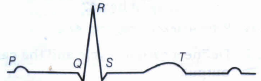
Given below is the diagrammatic representation of a standard ECG. Label its different peaks.
3.
Define systole and diastole.
4.
List the location of each of the valves of the heart.
5.
What are two heart sounds? When and how are these sounds produced?
6.
One of the ABO blood groups is sometimes called the universal donor. Which group do you think it is and why?
7.
What is the significance of time gap in the passage of action potential from sinoatrial node to the ventricle?
8.
What happens to the mitral valve and the related blood flow during ventricular systole?
9.
Sinoatrial node is called the pacemaker of our heart. Why?
10.
Where does the cardiac impulse originate?
11.
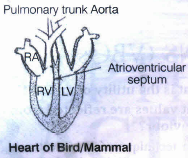
Explain the advantage of the complete partition of ventricle among birds and mammals and hence leading to double circulation.
12.
How can we say that artificial pacemaker is a life saving instrument?
13.
Explain the chemical events that takes place to form a blood clot to seal the wound.
14.
An Rh- woman is carrying an Rh+ foetus for the second time. Describe the consequences of Rh- incompatibility in this case?
15.
What is the significance of hepatic portal system in the circulatory system?
16.
Write the differences between :
(a) Blood and Lymph
(b) Open and Closed system of circulation
(c) Systole and Diastole
(d) P-wave and T-wave
17.
Why do we consider blood as a connective tissue?
18.
Explain different types of blood groups and donor compatibility making a table.
19.
Systole
20.
Platelets
21.
AB Group
22.
RBC
23.
Match column I with column II:
|
Column I |
Column II |
||
|
(a) |
Eosinophils |
(i) |
Coagulation |
|
(b) |
RBC |
(ii) |
Universal Recipient |
|
(c) |
AB Group |
(iii) |
Resist Infections |
|
(d) |
Platelets |
(iv) |
Contraction of Heart |
|
(e) |
Systole |
(v) |
Gas transport |
1.
The atrioventricular (AV) node is present in the right atrium, near the base of the septum that separates the right atrium from the ventricle. It gives rise to the bundle of His that conducts the cardiac impulses from the autrium to the ventricles. As the bundle of His passes the ventricle along the inter-ventricular septum, it divides into two branches – the right ventricle and the left ventricle.
The end branches of this conducting system then forms a network of Purkinje fibres that penetrate into the myocardium. The wave of excitation from the sino-atrial node (SA node) stimulates the atrio-ventricular node, thereby leading to the contraction of ventricles through the bundle of His and Purkinje fibres. Hence, the atrio-ventricular node and the atrioventricular bundle play a role in the contraction of ventricles.
2.
The representation of a standard ECG diagram

3.
Contraction of heart muscles brings right and left atria at systole and relaxation of heart muscles brings them at diastole.
4.
The tricuspid valves are present between right atrium and ventricle.Bicuspid valves are between the wall of left atrium and left ventricle. The semilunar valves are present in the inner wall of aorta.
5.
We can hear with a stethoscope, the two main heart sounds(i.e., lubb and dupp), which repeat rythmically. These sounds result from the closure of the heart valves. The first sound (lubb), which is of longer duration (0.16-0.90 s) and a louder one is created by the closure of the atrioventricular valves immediately after the start of the ventricular systole. The second sound is of shorter duration (0.10 s) and is created by the closure of the semilunar valve at the end of the ventricular systole.
6.
Individuals with blood group O are called universal donors. These individuals do not have either A or B antigen on the surface of their RBCs, but their blood serum contains antibodies both A and B antigens. Therefore, group individuals can only receive blood from a group O individual, but they can donate blood to individuals of any ABO blood group (i.e., A, B, O or AB).
7.
The time gap in the passage of action potential from sinoatrial node to the ventricles allows ventricles to relax. The ventricular pressure falls, causing the closure of semilunar valves, which prevents the backflow of blood into the ventricles.
8.
At the start of the ventricular systole, the mitral valve closes to prevent the flow of blood back into the atrium. As systole continues, blood from the right ventricle enters the pulmonary artery and blood from the left ventricle enters the aorta.
9.
SA node
1. The sino-atrial (SA) node is a specialized bundle of neurons located in the upper part of the right atrium of the heart.
2. The cardiac impulse originating from the SA node triggers a sequence of electrical events in the heart, thereby controlling the sequence of muscle contraction that pumps blood out of the heart.
3. Since the SA node initiates and maintains the rhythmicity of the heart, it is known as the natural pacemaker of the human body.
10.
The cardiac impulse originates in cardiac muscle fibres and is not brought to the heart by any nerve fibres. The origin of cardiac impulse is said to be myogenic.
11.
The birds and mammals have evolutionary advancement as far as structure of heart is concerned. They need more oxygen to live in terrestrial habitat.
In these animals, the blood received by left and right auricles is oxygenated and deoxygenated, respectively.
It passes towards the left and right ventricles and thus, no oxygenated and deoxygenated blood is mixed. The ventricles pump oxygenated and deoxygenated blood without mixing.
Thus, two separate circulatory pathways are found and this type of blood circulation is called as double circulation, which includes systemic and pulmonary circulation.
12.
It is an electronic device, which regularly sends a small amount of electrical charge for maintaining the rhythmicity of the heart. It is implanted subcutaneously in the upper thoracic region having a connection with the heart.
The artificial pacemaker is called a life-saving instrument because, it stimulates the heart electrically at regular intervals to maintain its beats. Thus, it replaces the SA node as the originator of the cardiac impluse. It is to be noted that some pacemakers stimulate the heart at a fixed rate, others sense when the natural heart rate falls below a predetermined value and then stimulates the heart.
13.
Chemical events that takes place from a blood clot are as follows
(i) When blood comes out of a blood vessel in case of injury the platelets clump together, break and release platelet factors, thromboplastin.
(ii) The enzyme thrombokinase initiates the conversion of prothrombin into thrombin.
(iii) Thrombin thus, catalysis the conversion of fibrinogen into fibrin which forms a mesh/network in which blood cells get entangled.
(iv) Ca2+ ions are necessary for both the above steps
14.
Rh-antigen is present on the surface of erythrocytes in about 80-85% of the human beings. The individuals, who possess this antigen are called Rh+ (Rh positive) and those, who do not have it are called Rh- (Rh negative). A person when exposed to Rh+ blood, develops anti Rh-antibodies. A pregnant women who is Rh-, if bears an Rh+ foetus, will develop anti-Rh-antibodies during the first delivery, when the foetal blood comes in contact with her blood. These antibodies linger in the blood for sufficiently longer periods. If she carries a second foetus, that is Rh+, the anti-Rh-antibodies in her blood enter the foetal circulation and cause damage to the foetal RBCs. This, could become fatal. This condition is called erythroblastosis foetalis.
15.
A unique vascular connection exists between the digestive tract and liver called hepatic poratal system.The hepatic portal vein carries blood from intestine to the liver before it is delivered to the systemic circulation.This ensures that the liver, which has the metabolic versatility to interconvert various organic molecules has first access to nutrients after the food is digested.
16.
(a) Blood contains RBCs and hence can transport gases. Lymph doesn't contain RBCs and cannot transport gases. Lymph mainly contains WBCs and play a role in the immune system of the body.
(b) The Open Circulatory System is a system in which fluid (called hemolymph) in a cavity called the hemocoel bathes the organs directly with oxygen and nutrients and there is no distinction between blood and interstitial fluid; this combined fluid is called hemolymph or haemolymph. The cardiovascular systems of humans are closed, meaning that the blood never leaves the network of blood vessels.
(c) Systole is the contraction of heart muscle and diastole is the dilatation of the heart muscle.
(d) Each peak in the ECG is identified with a letter from P to T that corresponds to a specific electrical activity of the heart. The P-wave represents the electrical excitation (or depolarisation) of the atria, which leads to the contraction of both the atria. The QRS complex represents the depolarisation of the ventricles, which initiates the ventricular contraction. The contraction starts shortly after Q and marks the beginning of the systole. The T-wave represents the return of the ventricles from excited to normal state (repolarisation). The end of the T-wave marks the end of systole.
17.
Connective Tissue: It is largely a category of exclusion rather than one with a precise definition, but all or most tissues in this category are similarly:
(a) Involved in structure and support.
(b) Derived from mesoderm, usually.
(c) Characterized largely by the traits of non-living tissue.
Blood is considered a connective tissue for two basic reasons:
(i) embryologically, it has the same origin (mesodermal) as do the other connective tissue types and
(ii) blood connects the body systems together bringing the needed oxygen, nutrients, hormones and other signaling molecules, and removing the wastes. As the name implies, connective tissue serves a "connecting" function. It supports and binds other tissues. Unlike epithelial tissue, connective tissue typically has cells scattered throughout an extracellular matrix. Interestingly enough, blood is considered to be a type of connective tissue. Even though it has a different function in comparison to other connective tissues it does have an extracellular matrix. The matrix is the plasma and erythrocytes, leucocytes and platelets are suspended in the plasma.
18.
Two groupings, i.e., the ABO and Rh- are widely used all over the world. ABO grouping is based on the presence or absence of two surface antigens (chemicals that can induce immune response) on the RBCs, i.e., A and B. Similarly, the plasma of different individuals contain two natural antibodies (proteins produced in response to antigens).
Blood Groups and Donor Compatibility
| Blood Group | Antigen on RCBs | Antibody in Plasma | Donor's Group |
| A | A | Anti-B | A, O |
| B | B | Anti-A | B, O |
| AB | A, B | Nil | AB, A, B, O |
| O | Nil | Anti- A, B | O |
From the above-mentioned table, it is evident that group 'O' blood can be donated to persons with any other blood group and hence 'O' group individuals are called 'universal donors'. Persons with 'AB' group can accept blood from persons with AB as well as the other groups of blood. Therefore, such persons are called 'universal recipients'.
19.
()
Contraction of Heart
20.
()
Coagulation
21.
()
Universal Recipient
22.
()
Gas Transport
23.
()
|
Column I |
Column II |
||
|
(a) |
Eosinophils |
(iii) |
Resist infections |
|
(b) |
RBC |
(v) |
Gas transport |
|
(c) |
AB Group |
(ii) |
Universal Recipient |
|
(d) |
Platelets |
(i) |
Coagulation |
|
(e) |
Systole |
(iv) |
Contraction of heart |

11th Standard CBSE Syllabus & Materials
11th Standard CBSE
CBSE 11th Business Studies Forms of Business Organisation Sample Question Papers Study Material - QB365 Set A
NEW11th Standard CBSE
CBSE 11th Business Studies Business, Trade and Commerce Sample Question Papers Study Material - QB365 Set A
NEW11th Standard CBSE
CBSE 11th Physics Waves Sample Question Papers Study Material - QB365 Set A
NEW11th Standard CBSE
CBSE 11th Physics Kinetic Theory Sample Question Papers Study Material - QB365 Set A

CBSE 11th Standard CBSE Subjects
CBSE Standards