 100% FREE ACCESS
11th Standard CBSE Syllabus & Materials
11th Standard CBSE
CBSE 11th Economics PART-A - Presentation of Data - New Sample Question Papers Study Material - QB365 Set A
NEW11th Standard CBSE
CBSE 11th Economics PART-A - Organisation of Data - New Sample Question Papers Study Material - QB365 Set A
NEW11th Standard CBSE
CBSE 11th Economics PART-A - Collection of Data - New Sample Question Papers Study Material - QB365 Set A
NEW11th Standard CBSE
CBSE 11th Economics PART-A - Introduction to Economics and Statistics - New Sample Question Papers Study Material - QB365 Set A
NEW11th Standard CBSE
CBSE 11th Business Studies International Trade Sample Question Papers Study Material - QB365 Set A
NEW11th Standard CBSE
CBSE 11th Business Studies Evolution and Fundamentals of Business Sample Question Papers Study Material - QB365 Set A

Published on: 12/10/2019
Locomotion and Movement
Download CBSE Class 11th Standard CBSE Biology question papers, sample papers, important questions, and previous year solved papers in PDF format. Get free study materials, NCERT solutions, and exam preparation resources for Class 11th Standard CBSE Biology
Questions + Answers key
Take MCQ Biology Test
1.
Describe different types of joints in the human skeleton.
2.
Explain the structure of contractile protein in skeletal muscle.
3.
Describe the structure of skeletal muscle.
4.
Write the difference between:
(a) Actin and Myosin
(b) Red and White muscles
(c) Pectoral and Pelvic girdle
5.
(i) How does a muscle return to its original form during relaxation?Also draw a diagram showing relaxation of muscle.
(ii) What causes muscle fatigue?
6.
Differentiate between pectoral and pelvic girdle
7.
What is osteoporosis? Why are women at a greater risk of suffering from osteoporosis? Suggest some precautionary means to avoid osteoporosis.
8.
Give a description of the appendicular skeleton in human.
9.
Describe the structure of the rib cage of human.
10.
Explain the structure of the vertebral column of human.
1.
Joints have been classified into three major structural forms, namely, fibrous, cartilaginous and synovial:
(a) Fibrous Joints :
Fibrous joints do not allow any movement. This type of joint is shown by the flat skull bones which fuse end-to-end with the help of dense fibrous connective tissues in the form of sutures, to form the cranium.
(b) Cartilaginous Joints :
In cartilaginous joints, the bones involved are joined together with the help of cartilages. The joint between the adjacent vertebrae in the vertebral column is of this pattern and it permits limited movements.
(c) Synovial Joints. Synovial joints are characterised by the presence of a fluid filled synovial cavity between the articulating surfaces of the two bones. Such an arrangement allows considerable movement. These joints help in locomotion and many other movements.
i) Ball and socket joint (between humerus and pectoral girdle),
ii) Hinge joint (knee joint),
iii) Pivot joint (between atlas and axis),
iv) Gliding joint (between the carpals) and
v) Saddle joint (between carpal and metacarpal of thumb).
2.
Structure of Contractile Proteins
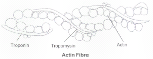
i) Each actin (thin) filament is made of two 'F' (filamentous) actins helically wound to each other. Each 'F' actin is a polymer of monomeric 'G' (Globular) actins.
ii) Two filaments of another protein, tropomyosin also run close to the 'F' actins throughout its length.
iii) A complex protein Troponin is distributed at regular intervals on the tropomyosin. In the resting state a subunit of troponin masks the active binding sites for myosin on the actin filaments.
Each myosin (thick) filament is also a polymerised protein. Many monomeric proteins called Meromyosins constitute one thick filament. Each meromyosin has two important parts, a globular head with a short arm and a tail, the former being called the heavy meromyosin (HMM) and the latter, the light meromyosin (LMM).
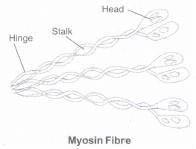
The HMI component, i.e.; the head and short arm projects outwards at regular distance and angle from each other from the surface of a polymerised myosin filament and is known as cross arm. The globular head is an active ATPase enzyme and has binding sites for ATP and active sites for actin.
3.
Structure of Skeletal Muscle
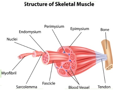
i) Each organised skeletal muscle is made of a number of muscle bundles or fascicles held together by a common collagenous connective tissue layer called fascia. Each muscle bundle contains a number of muscle fibres.
ii) Each muscle fibre is lined by the plasma membrane called sarcolemma enclosing the sarcoplasm. Muscle fibre is a syncitium as the sarcoplasm contains many nuclei.
iii) The endoplasmic reticulum, i.e., sarcoplasmic reticulum of the muscle fibres is the storehouse of calcium ions.
iv) A characteristic feature of the muscle fibre is the presence of a large number of parallelly arranged filaments in the sarcoplasm called myofilaments or myofibrils. Each myofibril has alternate dark and light bands on it.
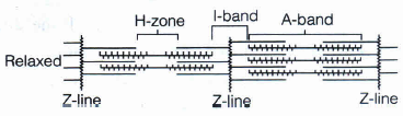
v) The striated appearance is due to the distribution pattern of two important proteins - Actin and Myosin. The light bands contain actin and is called I-band or Isotropic band, whereas the dark band called 'A' or Anisotropic band contains myosin. Both the proteins are arranged as rod-like structures, parallel to each other and also to the longitudinal axis of the myofibrils.
vi) Actin filaments are thinner as compared to the myosin filaments, hence are commonly called thin and thick filaments respectively.
vii) In the centre of each 'I' band is an elastic fibre called 'Z' line which bisects it. The thin filaments are firmly attached to the 'Z' line.
viii) The thick filaments in the 'A' band are also held together in the middle of this band by a thin fibrous membrane called 'M' line. The 'A' and 'I' bands are arranged alternately throughout the length of the myofibrils.
ix) The portion of the myofibril between two successive 'Z' lines is considered as the functional unit of contraction and is called a sarcomere.
x) In a resting state, the edges of thin filaments on either side of the thick filaments partially overlap the free ends of the thick filaments leaving the central part of the thick filaments. This central part of thick filament, not overlapped by thin filaments is called the 'H' zone.
4.
a) Actin and Myosin :
Actin filaments are thinner as compared to the myosin filaments, hence are commonly called thin and thick filaments respectively. Each actin (thin) filament is made of two 'F' (filamentous) actins helically wound to each other while each myosin (thick) filament is also a polymerised protein. Many monomeric proteins called Meromyosins constitute one thick filament.
(b) Red and White Muscles :
Muscle contains a red coloured oxygen storing pigment called myoglobin. Myoglobin content is high in some of the muscles which gives a reddish appearance. Such muscles are called the Red fibres. These muscles also contain plenty of mitochondria which can utilise the large amount of oxygen stored in them for ATP production. These muscles, therefore, can also be called aerobic muscles. On the other hand, some of the muscles possess very less quantity of myoglobin and therefore, appear pale or whitish. These are the White fibres. Number of mitochondria are also few in them, but the amount of sarcoplasmic reticulum is high. They depend on anaerobic process for energy.
(c) Pectoral and Pelvic Girdle :
Pectoral girdle is situated in the pectoral region of the body. Each half of pectoral girdle consists of a clavicleand a scapula. Scapula is a large triangular flat bone situated in the dorsal part of the thorax between the second and the seventh ribs. The dorsal, flat, triangular body of scapula has a slightly elevated ridge called the spine which projects as a flat, expanded process called the acromion. The clavicle articulates with this. Below the acromion is a depression called the glenoid cavity which articulates with the head of the humerus to form the shoulder joint. Each clavicle is a long slender bone with two curvatures. This bone is commonly called the collar bone. Pelvic girdle is situated in the pelvic region of the body. Pelvic girdle consists of two coxal bones. Each coxal bone is formed by the fusion of three bones - ilium, ischium and pubis. At the point of fusion of the above bones is a cavity called acetabulum to which the thigh bone articulates. The two halves of the pelvic girdle meet ventrally to form the pubic symphysis containing fibrous cartilage.
5.
(i) The calcium ions after contraction are quickly pumped back to the sarcoplasmic cisternae.This leads to blocking of active sites on actin.The Z-line returns to the original position.Causing muscles to return to its original position, i.e., relaxation form.
(ii) Repeated activation of the muscles leads to accumulation of lactic acid due to anaerobic breakdown of glycogen in them which causes fatigue.
6.
Pectoral and pelvic girdle help in the articulation of upper and lower limbs, respectively. Each girdle is made of two equal halves.
Each half of a pectoral girdle consists of clavicle and scapula. Scapula is a large triangular flat bone. There is glenoid cavity at the joint of scapula, clavicle and acromian process, which articulates with the head of humerus to form the shoulder joint.
Each half of pelvic girdle is formed by three bones, i.e., ilium, ischium and pubis. At the point of their fusion, there is a cavity called acetabulum to which the head of femur articulates.
7.
Osteoporosis is the condition in which the bone mass is decreased. This leads to general weakening of bones. Even a minor slip can result in fractures if a person is suffering from osteoporosis.
Women are at greater risk of suffering from osteoporosis because of several reasons. During child bearing major part of calcium from their body is utilized in formation of the skeleton of the foetus. Additionally during lactation there is further depletion of the calcium reserve of the body because of milk formation. A calcium rich diet can help prevent the progression of osteoporosis. Milk and eggs are good source of calcium. During pregnancy oral calcium supplement is also given to prevent osteoporosis.
8.
Appendicular Skeleton. The bones of the limbs alongwith their girdles constitute the appendicular skeleton. Each limb is made of 30 bones.
| Bones of Limbs | |
| Fore Limb | Hind Limb |
| Humerus, Radius, Ulna Carpals (8) Metacarpals (5) Phalanges (14) |
Femur, Tibia, Fibula, Tarsals (7) Metatarsals (5) Phalanges (14) Patella |
9.
Rib Cage. There are 12 pairs of ribs. Each rib is a thin flat bone connected dorsally to the vertebral column and ventrally to the sternum. It has two articulation surfaces on its dorsal end and is hence called bicephalic. First seven pairs of ribs are called true ribs. Dorsally, they are attached to the thoracic vertebrae and ventrally connected to the sternum with the help of hyaline cartilage. The 8th, 9th and 10th pairs of ribs do not articulate directly with the sternum but join the seventh rib with the help of hyaline cartilage. These are called vertebrochondral (false) ribs. Last 2 pairs (llth and 12th) of ribs are not connected ventrally and are therefore, called floating ribs. Thoracic vertebrae, ribs and sternum together form the rib cage.
10.
Vertebral Column. Our vertebral column is formed by 26 serially arranged units called vertebrae and is dorsally placed. It extends from the base of the skull and constitutes the main framework of the trunk. Each vertebra has a central hollow portion (neural canal) through which the spinal cord passes. First vertebra is the atlas and it articulates with the occipital condyles. The vertebral column is differentiated into followingregions starting from the skull:
1. cervical (7),
2. thoracic (12),
3. lumbar (5),
4. sacral (I-fused) and
5. coccygeal (I-fused] regions
The number of cervical vertebrae are seven in almost all mammals including human beings. The vertebral column protects the spinal cord, supports the head and serves as the point of attachment for the ribs and musculature of the back. Sternum is a flat bone on the ventral midline of thorax.

11th Standard CBSE Syllabus & Materials
11th Standard CBSE
CBSE 11th Business Studies Forms of Business Organisation Sample Question Papers Study Material - QB365 Set A
NEW11th Standard CBSE
CBSE 11th Business Studies Business, Trade and Commerce Sample Question Papers Study Material - QB365 Set A
NEW11th Standard CBSE
CBSE 11th Physics Waves Sample Question Papers Study Material - QB365 Set A
NEW11th Standard CBSE
CBSE 11th Physics Kinetic Theory Sample Question Papers Study Material - QB365 Set A



CBSE 11th Standard CBSE Subjects
CBSE Standards