 100% FREE ACCESS
12th Standard CBSE Syllabus & Materials
12th Standard CBSE
CBSE 12th Economics Government Budget and the Economy Previous year Question Papers Study Material - QB365 Set A
NEW12th Standard CBSE
CBSE 12th Computer Science Interface Python with MySQL - New Previous year Question Papers Study Material - QB365 Set A
NEW12th Standard CBSE
CBSE 12th Computer Science Database Concept - New Previous year Question Papers Study Material - QB365 Set A
NEW12th Standard CBSE
CBSE 12th Computer Science Data Communication - New Previous year Question Papers Study Material - QB365 Set A
NEW12th Standard CBSE
CBSE 12th Computer Science Data Structures - New Previous year Question Papers Study Material - QB365 Set A
NEW12th Standard CBSE
CBSE 12th Computer Science Functions - New Previous year Question Papers Study Material - QB365 Set A

Published on: 05/03/2026
Download CBSE Class 12th Standard CBSE Biology question papers, sample papers, important questions, and previous year solved papers in PDF format. Get free study materials, NCERT solutions, and exam preparation resources for Class 12th Standard CBSE Biology
Questions + Answers key
Take MCQ Biology Test
1.
Write a note on the development of endosperm. Mention the types with examples.
2.
What do you mean by development of embryo? Support the answer with diagram.
3.
With a neat, labelled diagram, describe the parts of a typical angiosperm ovule.
4.
Differentiate between microsporogenesis and megasporogenesis. Which type of cell division occurs during these events? Name the structures formed at the end of these two events.
5.
Explain with help of a diagram, the development of a mature embryo sac from a megaspore mother cell in an angiosperm.
6.
(a) Describe in sequence the process of megasporogenesis in angiosperms.
(b) Draw the seven-celled structure formed and label all the different cells.
7.
(a) Draw a labelled schematic diagram of the transverse section mature anther of an angiospermic plant.
(b) Describe the characteristic features of an insect-pollinated flower.
8.
(a) Describe the process of syngamy and triple fusion in angiosperms.
(b) Explain the development of a fertilised egg upto a mature embryo in a dicot plant. draw a labelled diagram of a mature dicot embryo,
9.
(a) Differentiate between: Autogamy, geitonogamy and xenogamy.
(b) Explain the events that occur during pollen-pistil interaction.
10.
How does a pollen mother cell develop into a mature pollen grain? Illustrate the stages with labelled diagrams.
1.
Development of endosperm. Endosperm develops after double fertilization in angiosperms. It is triploid and develops from primary endosperm nucleus. In angiosperms three types of endosperm development are observed:
(1) Nuclear endosperm. In nuclear endosperm development nucleus divides by free nuclear division, there is appearance of central vacuole, all the nuclei are pushed towards periphery then starts cytokinesis and results into the formation of cells hut in coconut vacuole persists which is filled with fluid.
(2) Cellular endosperm. In cellular endosperm each and every division is followed by cytokinesis, so endosperm is cellular from the very beginning e.g. Datura, Petunia.
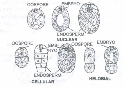
(3) Helobial endosperm. It is intermediate between cellular and nuclear endosperm. The first division is followed by cytokinesis to form two unequal cells, micropylar cell and chalazal cell. Further divisions in each cell are free nuclear divisions. This type of endosperm is found in order Helobiales.
2.
(a) (i) Endosperm development precedes embryo development. The primary endosperm cells divide repeatedly and forms a triploid endosperm tissue. The cells of this tissue are filled with reserve food materials and are used for development. (ii) Endosperm persists in albuminous seeds (castor and coconut). Endosperm are used during seed germination.
(b) Development of embryo. 1. The zygote or oospore divides by a transverse wall into an upper suspensor cell and lower embryonal cell.
2. The suspensor cell which lies towards the micropylar end, divides by transverse divisions to constitute 7-10 cells called suspensor.
3. The upper cell of the suspensor filament towards micropylar end is called haustorial cell, whereas the celllying above the embryo cell is called hypophysis.
4. The haustorial cell enlarges in size and attaches the suspensor to the tip of embryo sac.
5.The embryonal cell divides by second longitudinal division at right angle to the first and then by transverse division to form an octant or eight celled embryo.
6. Out of these eight cells, the lower four cells of octant away from the suspensor give rise to the plumule and the two cotyledons, while the above four cells of octant near the suspensor forms the hypocotyl and stele of radicle.
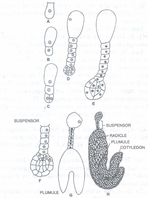
7. Now this octant divides by a periclinal division to form outer single layered dermatogen from which arises the epidermis layer. The inner cells further divide to form periblem below the dermatogen and the central plerome.
8. The periblem forms the cortex while the plerome gives stele of embryo.
9. The lowermost cell of suspensor, which is lying just above the octant cells is known as hypophysis. The hypophysis divides to give rise the dermatogen and periblem of the radicle.
10. A fully developed embryo of dicotyledons has an embryonal axis differentiated into plumule, two cotyledons and radicle.
11.In the beginning embryo is globular. When two cotyledons differentiate from the sides with faint plumule in the centre, embryo become heart-shaped. Now the embryo undergoes rest and ovule is transformed into seed.
Monocot embryo. Unlike the dicots, where the embryonal mass is formed of eight cells, the anterior cells forming the plumule and the cotyledons, and the posterior forming hypocotyl, the development is much variable in monocots i.e. grass family. In some cases, suspensor does not develop at all. Only one cotyledon appears in monocots as a terminal structure. The plumule always appears laterally from it.
3.
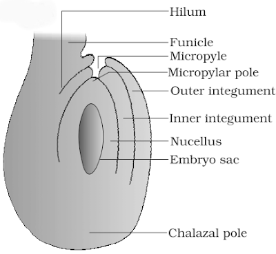
1. The ovule is attached to the placenta by a short stalk called funiculus.
2. The junction between the funiculus and the ovule, is called hilum.
3. The primordium of the ovule grows into a mass of cells, forming the nucellus, the main body of the ovule.
4. It becomes surrounded by two protective envelopes called integuments, which leave a small opening called micropyle, at the tip of the ovule.
5. The basal part of the ovule (opposite to the micropyle) is called chalaza. Cells of the nucellus are rich in reserve food materials.
6. There is a single embryo sac/female gametophyte in the nucellus; it has developed from a megaspore.
7. The embryo sac contains the following cells:
(i) Three antipodal cells at the chalazal end.
(ii) Three cells at the micropylar end, forming egg apparatus, which consists of two synergids and a female gamete.
(iii) Two nuclei in the central cell, called polar nuclei, which fuse to form a diploid secondary nucleus.
4.
|
Microsporogenesis |
Megasporogenesis |
|
It occurs inside the microsporangia of anther. |
It occurs in the nucellus of the ovule/megasporangium. |
Meiosis occurs during sporogenesis.
The structure formed at the end are microspores and megaspores respectively.
5.
Development of Female Gametophyte:
- The megaspore mother cell undergoes meiosis to form four megaspores.
- Three of them degenerate and the functional megaspore enlarges to form the embryo sac.
- Its haploid nucleus undergoes a mitotic division and the two daughter nuclei move towards the poles.
- Each of them undergoes two successive mitotic divisions.
- Cell wall formation starts in the 8-nucleate stage.
- Three of them at the micropylar end form the egg apparatus, consisting of a female gamete and two synergids.
- Three of them at the chalazal end form the antipodal cells.
- One nucleus from each of the poles moves to the centre; they are called polar nuclei.
- The two polar nuclei fuse in the centre (now the central cell) to form a diploid secondary nucleus.
- Thus, the mature embryo sac is 7-celled and 8-nucleate.
6.
Development of Female Gametophyte:
- The megaspore mother cell undergoes meiosis to form four megaspores.
- Three of them degenerate and the functional megaspore enlarges to form the embryo sac.
- Its haploid nucleus undergoes a mitotic division and the two daughter nuclei move towards the poles.
- Each of them undergoes two successive mitotic divisions.
- Cell wall formation starts in the 8-nucleate stage.
- Three of them at the micropylar end form the egg apparatus, consisting of a female gamete and two synergids.
- Three of them at the chalazal end form the antipodal cells.
- One nucleus from each of the poles moves to the centre; they are called polar nuclei.
- The two polar nuclei fuse in the centre (now the central cell) to form a diploid secondary nucleus.
- Thus, the mature embryo sac is 7-celled and 8-nucleate.
7.
(i) Insect-pollinated flowers are large, colourful and showy; if flowers are small, they are clustered into inflorescene to make them conspicuous.
(ii) The pollen grains and stigmatic surface are sticky.
(iii) They are highly fragrant; flowers pollinated by flies and beetle have foul odours to attract them.
(iv) To sustain animal visits, the flowers offer certain rewards like nectar, edible pollen and safe place for laying eggs.
8.
(a) Syngamy
Syngamy refers to the fusion of a male gamete with the female gamete in the embryo sac to form a diploid zygote.
Triple fusion
Triple fusion refers to the fusion of a male gamete with the secondary nucleus (formed by fusion of two haploid polar nuclei) to form the triploid primary endosperm nucleus.
(b) Embryo development
Th'e zygote undergoes mitotic divisions during embryogeny.
The first division is transverse and results in a basal cell and a terminal cell; the basal cell forms the suspensor while the terminal cell forms the embryo proper.
It first, forms a filamentous proembryo, which later forms in sequence a globular embryo, heart-shaped embryo and then a mature embryo.
9.
(a) Differences:
| Autogamy | Getonogamy | Xenogamy |
| It refers to the transfer of pollen grains from the anther of a flower to the stigma of the same flower | It refers to the transfer of pollen grains from the anther of a flower to the stigma of same plant. | It refers to the transfer of pollen grains from the anther of a flower to the stigma of another flower on a different plant of the same species. |
| It does not lead to genetic variation in the progency | It does not lead to genetic variation in the progeny. | It leads to genetic variation in the progeny |
| No pollinating agent is needed. | A pollinating agent is needed. | A pollinating agent is needed. |
(b) Pollen-Pistil Interaction.
The stigma/pistil has the ability to recognise the right type of pollen, i.e. the compatible pollen of the same species and rejects the wrong type of pollen grains.
It is the result of interaction between the chemical components of the pollen and those of stigma. - A compatible pollen grain starts its germination on the stigma.
The intine grows out through one of the germ pores as a protuberance.
The contents of the pollen move into this tube, i.e. the tube nucleus (nucleus of the vegetative cell) and the generative cell (or the two male gametes in those species where pollen is liberated in the three-celled stage).
The pollen tube grows through the tissues of stigma and style and enters the ovule through micropyle.
It enters the embryo sac through the filiform apparatus of one of the synergids to liberate the male gametes.
The events from the deposition of pollen on the stigma till the pollen tube enters the ovule, are collectively referred to as pollen-pistil interaction.
10.
(a) Development of Pollen grain:
- Microsporocytes undergo mitotic divisions to increase the number of functional microspore mother cells.
- Each microspore mother cell undergoes meiosis to form a microspore tetrad.
- All the four microspores remain enclosed in a common wall.
- They start separating from each other.
- The nucleus undergoes a mitotic division resulting in the formation of a large vegetative cell and a small generative cell.
- The spore develops a wall of two layers the outer- exine and inner intine.
- At this stage, the pollen grains are liberated; but in some cereals the generative cell undergoes mitosis and the pollen grain is 3-celled, at the time of liberation.
(b) Mature Pollen Grain:
- A mature pollen grain has a protective covering of two layers.
- The outer is thick, sculptured variously and is made of sporopollenin; this layer is called exine.
- The inner layer, called intine is thin and is made of cellulose.
- A mature pollen grain is two celled with a large vegetative cell and a small generative cell floating in the cytoplasm of the vegetative cell.
- The pollen grains of dicot plants have three germ pores while those of monocots have one.

12th Standard CBSE Syllabus & Materials
12th Standard CBSE
CBSE 12th Computer Science Python Revision Tour I - New Previous year Question Papers Study Material - QB365 Set A
NEW12th Standard CBSE
CBSE 12th Business Studies Planning Important Questions And Answers Study Material - QB365 Set A
NEW12th Standard CBSE
CBSE 12th Business Studies Business Environment Important Questions And Answers Study Material - QB365 Set A
NEW12th Standard CBSE
CBSE 12th Business Studies Principles of Management Important Questions And Answers Study Material - QB365 Set A



NCERT Books

Syllabus

Exam Pattern

Sample Question Papers

Previous year Question Papers

Important Notes

MCQ Practice test

NCERT Exemplers

Case study Questions

Image Based Questions

Passage based Questions

HOT Questions

Value Based Questions

Model Questions Papers
NCERT ( Book Back ) Questions

Assertion and Reason
Important Questions And Answers

CBSE 12th Standard CBSE Subjects
CBSE Standards