 100% FREE ACCESS
11th Standard Syllabus & Materials
11th Standard
TN 11th Tamil இயற்கை வேளாண்மை,சுற்றுச்சூழல் -செய்யுள் - மனோன்மணீயம் Important Questions And Answers Study Material - QB365 Set A
NEW11th Standard
TN 11th Tamil என்னுயிர் என்பேன் -துணைப்பாடம் - இசைத்தமிழர் இருவர் Important Questions And Answers Study Material - QB365 Set A
NEW11th Standard
TN 11th Tamil மொழி கலை -செய்யுள் - ஒவ்வொரு புல்லையும் Important Questions And Answers Study Material - QB365 Set A
NEW11th Standard
TN 11th Tamil பீடு பெற நில் - இலக்கணம் - பகுபத உறுப்புகள் Important Questions And Answers Study Material - QB365 Set A
NEW11th Standard
TN 11th Tamil பீடு பெற நில் - துணைப்பாடம் - வாடிவாசல் Important Questions And Answers Study Material - QB365 Set A
NEW11th Standard
TN 11th Tamil பீடு பெற நில் - செய்யுள் - குறுந்தொகை Important Questions And Answers Study Material - QB365 Set A
Published on: 12/03/2019
11th Public Exam March 2019 Important 5 Marks Questions
Download Tamil Nadu 11th Standard Biology question papers, model tests, one-mark questions, important questions, and public exam papers in PDF format. Free study materials and answer keys for TN State Board students.
Questions + Answers key
Take MCQ Biology Test1.
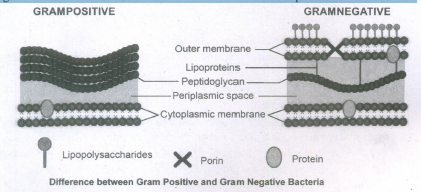
Describe the structure of Gram positive and Gram negative bacterial cell wall using diagram.
2.
Describe the Competitive & Non-Competitive Inhibitors of enzyme.
3.
List out the important features of Chromosomes.
4.
Define bud. Explain the types of buds based on location.
5.
Describe the Concept of species - Morphological, Biological and Phylogenetic
6.
Expalin the Fusion of stamens
7.
Describe the structure of Vacuoles
8.
General Characteristic features of Algae.
9.
Enumerate the differences between mitosis and meiosis.
10.
11.
Explain the reactions taking place in mitochondrial inner membrane.
12.
A transverse section of the trunk of a tree shows concentric rings which are known as growth rings. How are these rings formed? What are the significance of these rings?
13.
Distinguish the anatomy of dicot root from monocot root.
14.
Explain sclereids with their types.
15.
16.
Briefly explain the structure of thyroid gland.
17.
Describe the structures of olfactory receptors.
18.
Explain the sliding- filament theory of muscle contraction.
19.
What are the bones that make the pelvic girdle?
20.
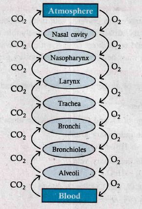
Sketch a flow chart to show the path way of air flow during respiration.
21.
Write a note on Haemodialysis.
22.
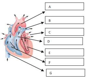
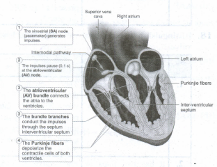
Name and Label the given diagrams to show A, B, C, D, E, F, and G
23.
24.
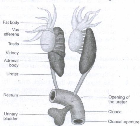
Explain the excretory system of frog.
25.
How respiration takes place in cockroach?
26.
Write the classification of connective tissue and their functions.
27.
List the salient features of phylum Annelida.
28.
Explain the role of Latin and Greek names in Biology.
29.
Write a note on multiple / Composite fruits.
30.
Write a note on DNA Barcoding.
31.
Describe the structure of Mitochondria.
32.
What are the different types of phyllotaxy?
33.
What are Mycorrhizae? Explain the types.
34.
Describe the Fine Structure of a cell wall.
35.
Describe the structure of Tobacco Mosaic Virus.
36.
List out the Economic importance of Gymnosperms.
37.
Explain the different types of fleshy fruit with suitable example.
38.
Write the characteristic features of DNA.
39.
Give a general account on lichens.
40.
What are the factors affecting the rate of enzyme reaction?
41.
Give the floral characters of Clitoria ternatea.
42.
43.
Write about Pachytene and Diplotene of Prophase I.
44.
What is tubular secretion? Name the substances secreted through the renal tubules.
45.
How are the kidneys involved in controlling blood volume? How is the volume of blood in the body related to arterial pressure?
46.
What is the difference between a Zoo and Wild Life Sanctuary?
47.
48.
How will you calculate net products of one sucrose molecule upon complete oxidation during aerobic respiration as per recent view?
49.
Give the economic importance of Silkworm
50.
Could the number of eggs or young ones produced by an oviparous and viviparous female be equal? Why?
1.
Most of the gram positive cell wall contain considerable amount of teichoic acid and teichuronic acid. In addition, they may contain polysaccharide molecules. The gram negative cell wall contains three components that lie outside the peptidoglycan layer:
1. Lipoprotein
2. Outer membrane and
3. Lipopolysaccharige.
Thus the different results in the gram stain are due to differences in the structure and composition of the cell wall.
2.
Competitive Inhibitor: Molecules that resemble the shape of the substrate and may compete to occupy the active site of enzyme are known as competitive inhibitors. For Example: the enzyme that catalyses the reaction between carbon dioxide and the CO2 acceptor molecule in photosynthesis, known as ribulose biphosphate carboxylase oxygenase (RUBISCO) is competitively inhibited by oxygen/carbon-di-oxide in the chloroplast. The competitive inhibitor is malonate for succinic dehydrogenase.
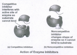
Non-competitive Inhibitors: There are certain inhibitors which may be unlike the substrate molecule but still combines with the enzyme. This either blocks the attachment of the substrate to active site or change the shape so that it is unable to accept the substrate. For example the effect of the amino acids alanine on the enzyme pyruvate kinase in the final step of glycolysis. Certain non-reversible/irreversible inhibitors bind tightly and permanently to an enzyme and destroy its catalytic properties entirely. These could also be termed as poisons. Example - cyanide ions which blocks cytochrome oxidase in terminal oxidation in cell aerobic respiration, the nerve gas sarin blocks a neurotransmitter in synapse transmission.
3.
The four important features of the chromosome are:
The shape of the chromosome is specific: The long, thin, lengthy structured chromosome contains a short, constricted region called centromere. A centromere may occur any where along the chromosome, but it is always in the same position -on any given chromosome. The number of chromosomes perspecies is fixed: For example the mouse has 40 chromosomes, the onion has 16 and humans have 46.
Chromosomes occur in pairs: The chromosomes of a cell occur in pairs, called homologous pairs. One of each pair come originally from each parent. Example, human has 46 chromosomes, 23 coming originally from each parent in the process of sexual reproduction. Chromosomes are copied: Between nuclear divisions, whilst the chromosomes are uncoiled and cannot be seen, each chromosome is copied. The two identical structures formed are called chromatids.
4.
Buds are the growing points surrounded by protective scale leaves.
1. Terminal bud or Apical bud: These buds are present at the apex of the main stem and at the tips of the branches.
2. Lateral bud or Axillary bud: These buds occur in the axil of the leaves and develop into a branch or flower.
3. Extra axillary bud: These buds are formed at nodes but outside the axil of the leaf as in Solanum americanum.
4. Accessory bud: An extra bud on either side (collateral bud) or above (superposed bud or serial bud) the axillary bud. e.g., Citrus and Duranta.
5.
Species is the fundamental unit of taxonomic classification. Greek philosopher Plato proposed concept of "eidos" or species and believed that all objects are shadows of the "eidos". Accordl[lg to Stebbins (1977) species is the basic unit of evolutionarv process. Species is a group of individual organisms which have the following characters.
1. A population of organisms which closely resemble each other more than the other population.
2. They descend from a common ancestor.
3. In sexually reproducing organisms, they interbreed freely in nature; producing fertile offspring.
4. In asexually reproducing organisms, they are identified by their morphological resemblance.
5. In case of fossil organisms, they are identified by the morphological and anatomical resemblance.
Species concepts can be classified into two general groups. Concept emphasizing process of evolution that maintains the species as a unit and that can result in evolutionary divergence and speciation: Another concept emphasises the product of evolution in defining a species.
Types of Species
There are different types of species and they are as follows:
1. Process of evolution - Biological Species
2. Product of evolution - Morphological Species and Phylogenetic Species
Morphological Species (Taxonomic species) When the individuals are similar to one another in one or more features and different from other such groups, they are called morphological species. These species are defined and categorized with no knowledge of phylogenetic history, gene flow or detailed reproductive mechanisms.
Biological Species (Isolation Species) According to Ernest Mayr 1963," these are groups of populations that interbreed and are reproductively isolated from other such groups in nature".
Phylogenetic Species
This concept was developed by Meglitsch (1954), Simpson (1961) and Wiley (1978). Wiley defined phylogenetic species as "an evolutionary species is a single lineage of ancestor descendent populations which maintains its identity from other such lineages which has its own evolutionary tendencies and historical fate".
6.
Refers to the stamens fusing among themselves or with other parts of flower. Two types.
Connation
Adnation
Connation:
Refers to the fusion of stamens among themselves. It is of 3 types.
a. Adelphy.
b. Syngenecious.
c. Synandrous.
Adelphy:
Filaments connate into one or more bundles but anthers are free. It may be the following types.
1. Monadelphous: Filaments of stamens connate into a single bundle. Example: Malvaceae (chinarose, cotton).
2. Diadelphous: Filaments of stamens connate: into two bundles. Example: Fabaceae, pea.
3. Polyadelphous: Filaments connate into many bundles. Example: Citrus, Bombax
Syngenesious: Anthers connate, filaments free. Example: Asteraceae.
Synandrous: Filaments and anthers are completely fused. Example: Coccinea.
Adnation: Refers to the fusion of stamens with other floral parts. Epipetalous (petalostemonous): Stamens are adnate to petals. Example: brinjal, Datura.
Episepalous: stamens are adnate to sepals. Example: Grevillea (Silver oak)
Epitepalous (epiphyllous): stamens are adnate .to tepa Is. Example: Aphodelus, Asparagus
Gynostegium:
Connation product of stamens and stigma is called gynostegium. Example: Calotropis and Orchidaceae.
Pollinium: Pollen grains are fusedtogether as a single mass.
7.
In plant cells vacuoles are large, bounded by a single unit membrane called Tonoplast. The vacuoles contain cell sap, which is a solution of sugars, amino acids, mineral salts, waste chemical and anthocyanin pigments. Beetroot cells contains anthocyanin pigments in their vacuoles. Vacuoles accumulate products like tannins. The osmotic expansion of a cell kept in water is chiefly regulated by vacuole and the water enters the vacuoles by osmosis.
The major function of plant vacuole is to maintain water pressure known as turgor pressure, which maintains the plant structure. Vacuoles organises itself into a storage/sequestration compartment. Example: Vacuoles store, most of the sucrose of the cell.
i. Sugar in Sugar beet and Sugar cane.
ii. Malic acid in Apple.
iii. Acids in Citrus fruits.
iv. Flavonoid pigment Cyanidin3 rutinoside in the petals of Antirrhinum.
v. Tannins in Mimosa pudica.
vi. Raphide crystals in Dieffenbachia.
vii. Heavy metals in Mustard (Brassica).
viii. Latex in Rubber tree and Dandelionstem.
8.
(i) The algae show a great diversity in size, shape and structure.
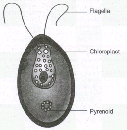
(ii) A wide range of thallus organisation is found in algae. Unicellular motile (Chlamydomonas), unicellular non-motile (Chlorella), Colonial motile (Volvox), Colonial non motile (Hydrodictyon), siphonous (Vaucheria), unbranched filamentous (Spirogyra), branched filamentous (Cladophora), discoid (Coleochaete) heterotrichous (Fritschiella), Foliaceous (Ulva) to Giant Kelps (Laminaria and Macrocystis).
(iii) The thallus organization in algae is given in Algae are Eukaryotes except blue green algae.
(iv) The plant body does not show differentiation into tissue systems.
(v) The cell wall of algae is made up of cellulose and hemicellulose.
(vi) Siliceous walls are present in diatoms. In Chara the thallus is encrusted with calcium carbonate. Some algae possess algin, polysulphate esters of polysaccharides which are the sources for the alginate, agar agar and Carrageenan.
(vii) The cell has a membrane bound nucleus and cell organelles like chloroplast, mitochondria, endoplasmic reticulum, golgi bodies etc., Pyrenoids are present.
(viii) They are proteinaceous bodies found in chromatophores and assist in the synthesis and storage of starch.
(ix) The pigmentation, reserve food material and flagellation differ among the algal groups.
(x) Algae reproduces by vegetative, asexual an sexual methods.
(xi) Vegetative reproduction includes fission (In unicellular forms the cell divides mitotically to produce two daughter cells.
(xii) (Example: Chlamydomonas); Fragmentation (fragments of parent thallus grow into new individual Example: Ulothrix) Budding (A lateral bud is formed in some members like Protosiphon and helps in reproduction) Bulbils, (a wedge shaped modified branch develop in Sphacelaria) Akinetes (Thick walled spores meant for perennation and germinates with the advent of favourable condition
(xiii) (Example: Pithophora). Tubers (Structures found on the rhizoids and the lower nodes of Chara which store food materials). Asexual reproduction takes place by the production of zoospores (Ulothrix, Oedogonium) aplanospore(thin walled non motile spores Exmaple: Vaucheria);
(xvi) Autospores (spores which look similar to parent cell Example: Chlorella); Hypnospore (thick walled aplanospore - Example: Chlamydomonas nilvalis); Tetraspores (Diploid thallus of Polysiphonia produce haploid spores after meiosis).
Sexual reproduction in algae are of three type
1. Isogamy (Fusion of morphologically and Physiologically similar gametes Example: Ulothrix)
2. Anisogamy (Fusion of either morphologically or physiologically dissimilar gametes Example: Pandorina)
3. Oogamy (Fusion of both morphologically and physiologically dissimilar gametes .Example: Sargassum).
The life Cycle shows distinct alternation of generation.
9.
| Mitosis | Meiosis |
| One division | Two divisions |
| Homologous chromosomes line up separately on the metaphase plate. | Homologous chromosomes line up in pairs at the metaphase plate |
| Homologous chromosome do not pair up | Homologous chromosome pair up to form bivalent. |
| Chiasmata do not form and crossing over never occurs. | Chiasmata form and crossing over occurs. |
| Daughter cells are genetically identical. | Daughter cells are genetically different from the parent cells. |
| Two daughter cells are formed | Four daughter cells are formed. |
10.
11.
Electron Transport Chain (ETC) (Terminal oxidation)
(i) During glycolysis, link reaction and Krebs cycle the respiratory substrates are oxidized at several steps and as a result many reduced coenzymes NADH + H+ and FADH2 are produced.
(ii) These reduced coenzymes are transported to inner membrane of mitochondria and are converted back to their oxidized forms produce electrons and protons.
(iii) In mitochondria, the inner membrane is folded in the form of finger projections towards the matrix called cristae.
(iv) In cristae many oxysomes (F1 particles) are present which have electron transport carriers are present.
(v) According to Peter Mitchell's Chemiosmotic theory this electron transport is coupled to ATP synthesis. Electron and hydrogen(proton) transport takes place across four multiprotein complexes(I-IV). They are:
a) Complex-I (NADH dehydrogenase)
(i) It contains a flavoprotein(FMN) and associated with non-heme iron Sulphur protein (Fe-S). This complex is responsible for passing electrons and protons from mitochondrial NADH (Internal) to Ubiquinone(UQ).
\(\mathrm{NADH}+\mathrm{H}^{+}+\mathrm{UQ} \rightleftharpoons \mathrm{NAD}^{+}+\mathrm{UQH}_{2}\)
(ii) In plants, an additional NADH dehydrogenase (External) complex is present on the outer surface of inner membrane of mitochondria which can oxidise cytosolic NADH + H+.
(iii) Because mitochondrial inner membrane cannot allow NADH molecules directly in to the matrix. Ubiquinone (UQ) or Coenzyme Quinone (CoQ) is a small, lipid soluble electron, proton carrier located within the inner membrane of mitochondria.
b) Complex-II (Succinic dehydrogenase)
(i) It contains FAD flavoprotein is associated with non-heme iron Sulphur (Fe-S) protein.
(ii) This complex receives electrons and protons from succinate in Krebs cycle and is converted into fumarate and passes to ubiquinone.

c) Complex-III (Cytochrome bc1 complex)
(i) This complex oxidises reduced ubiquinone (ubiquinol) and transfers the electrons through Cytochrome bc1 Complex (Iron Sulphur center bc1 complex) to cytochrome c.
(ii) Cytochrome c is a small protein attached to the outer surface of inner membrane and act as a mobile carrier to transfer electrons between complex III to complex IV.
\(\mathrm{UQH}_{2}+2 \mathrm{CytC}_{\text {oxidised }} \rightleftharpoons \mathrm{UQ}+2 \mathrm{Cytc}_{\text {reduced }}+2 \mathrm{H}^{+}\)
d) Complex IV (Cytochrome c oxidase)
(i) This complex contains two copper centers (A and B) and cytochromes a and a3. Complex IV is the terminal oxidase and brings about the reduction of 1/2 O2 to H2O.Two protons are needed to form a molecule of H2O (terminal oxidation).
\(2 \mathrm{CytC}_{\text {oxidised }}+2 \mathrm{H}^{+}+1 / 2 \mathrm{O}_{2} \rightleftharpoons 2 \mathrm{CytC}_{\text {reduced }}+\mathrm{H}_{2} \mathrm{O}\)
(ii) The transfer of electrons from reduced coenzyme NADH to oxygen via complexes I to IV is coupled to the synthesis of ATP from ADP and inorganic phosphate (Pi) which is called Oxidative phosphorylation.
(iii) The F0F1- ATP synthase (also called complex V) consists of F0 and F1. F1 converts ADP and Pi to ATP and is attached to the matrix side of the inner membrane.
(iv) F0 is present in inner membrane and acts as a channel through which protons come into matrix.
12.
Annual Rings
(i) The activity of Vascular cambium is under the control of many physiological and environmental factors.
(ii) In temperate regions, the climatic conditions are not uniform throughout the year.
(iiii) In the spring season, cambium is very active and produces a large number of xylary elements having Vessels/tracheids with wide lumen.
(iv) The wood formed during this season is called spring wood or early wood.
(v) The tracheary elements are fairly thin walled.
(vi) In winter, the cambium is less active and forms fewer xylary elements that have narrow vessels tracheids and this wood is called autumn wood or late wood.
(vii) The tracheary elements are with narrow lumen, very thick walled.
(viii) Spring wood is lighter in colour with lower density.
(ix) Autumn wood is darker and has higher density.
(x) Sometimes Annual rings are called growth rings but all growth rings are not annual rings.
(xi) In some trees more than one growth ring is formed within a year due to climatic change.
(xii) Additional growth rings are developed within a year due to adverse natural calamities like drought, frost, defoliation, flood, mechanical injury and biotic factors during the middle of a growing season, which results in the formation of more than one annual ring. Such rings are called pseudo or false-annual rings.
(xiii) Each annual ring corresponds to one year's growth and on the basis of these rings, the age of a particular plant can easily be calculated.
(xiv) The determination of the age of a tree by counting the annual rings is called dendrochronology.
13.
| S.No. | Characters | Dicot root | Monocot root |
|---|---|---|---|
| 1. | Pericyle | Gives rise to lateral roots, phellogen and a part of vascular cambium. | Gives rise to lateral roots only. |
| 2. | Vascular tissue | Usually limited number of xylem and phloem strips. | Usually more number of xylem and phloem strips. |
| 3. | Conjunctive tissue | Parenchymatous; Its cells are differentiated into vascular cambium. | Mostly sclerenchymatous but sometimes parenchymatous. It is never differentiated in to vascular cambium. |
| 4. | Cambium | It appears as a secondary meristem at the time of secondary growth. | It is altogether absent. |
| 5. | Xylem | Usually tetrach | Usually polyarch |
14.
Sclereids (Stone Cells)
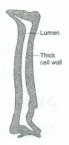
Sclereids are dead cells, usually these are isodiametric but some are elongated too. The cell wall is very thick due to lignification. Lumen is very much reduced. The pits may simple or branched. Sclereids are mechanical in function. They give hard texture to the seed coats, endosperms etc., Sclereids are classified into the folowing types.
Types of Sclereids
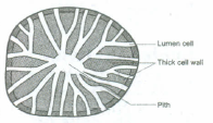
(a) Brachysclereids or Stone cells:
Isodiametric sclereids, with hard cell wall. It is found in bark, pith cortex, hard endosperm and fleshy portion of some fruits. Example: Pulp of Pyrus.
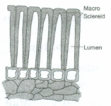
(b) Macrosclereids:
Elongated and rod shaped cells, found in the outer seed coat of leguminous plants. Example: Crotalaria and Pisum sativum.
(c) Osteosclereids (Bone cells):
Rod shaped with dilated ends. They occur in leaves and seed coats. Example: seed coat of Pisum and Hakea
(d) Astrosclereids:
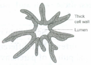
Star cells with lobes or arms diverging form a central body. They occur in petioles and leaves. Ex: Tea, Nymphae and Trochodendron.
(e) Trichosclereids:
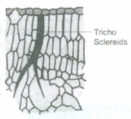
Hair like thin walled sclereids. Numerous small angular crystals are embedded in the wall of these sclereids, present in stems and leaves of hydrophytes. Example: Nymphaea leaf and Aerial roots of Monstera.
15.
16.
Structure of thyroid gland:
Thyroid gland is the largest endocrine gland in the body.
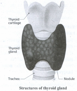
1. It is a butterfly shaped, bilobed gland located below the larynx on either side of upper trachea.
2. The two lateral lobes are connected by a median tissue mass called isthmus.
3. Each lobe is made up of many lobules. The lobules consist of follicles called acini.
4. Each acinus is lined with glandular, cuboidal or squamous epithelial cells. The lumen of acinus is filled with colloid, a thick glycoprotein mixture consisting of thyroglobulin molecules.
5. It secretes two hormones namely tri-iodothyronine [T3] and thyroxine or tetra- iodothyronine [T4].
6. The parafollicular cells or 'C' cells of thyroid gland secrete a hormone called thyrocalcitonin.
7. Iodine is essential for the normal synthesis of thyroid hormone.
8. Thyroid hormones shows a negative feedback effect on the hypothalamus and pituitary.
17.
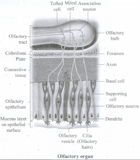
(i) The structure of chemoreceptors
(ii) The receptors for taste and smell are the chemoreceptors
(iii) The smell receptors are excited by air borne chemicals that dissolve in fluids.
(iv) The yellow coloured patches of olfactory epithelium form the olfactory organs that are located on the roof of the nasal cavity.
18.
Mechanism of muscle contraction
(i) Sliding filament theory in 1954, Andrew F. Huxley and Rolf Niedergerke proposed the sliding-filament theory to explain muscle contraction.
(ii) According to this theory, overlapping actin and myosin filaments of fixed length slide past one another in an energy requiring process, resulting in muscle contraction.
(iii) The contraction of muscle fibre is a remarkable process that helps in creating a force to move or to resist a load.
(iv) The force which is created by the contracting muscle is called muscle tension.
(v) The load is a weight or force that opposes contraction of a muscle.
(vi) Contraction is the creation of tension in the muscle which is an active process and relaxation is the release of tension created by contraction.
(vii) Muscle contraction is initiated by a nerve impulse sent by the central nervous system (CNS) through a motor neuron.
(viii) The junction between the motor neuron and the sarcolemma of the muscle fibre is called the neuromuscular junction or motor end plate.
(ix) When nerve impulse reaches a neuromuscular junction, acetylcholine is released.
(x) It initiates the opening of multiple gated channels in sarcolemma. The action potential travels along the T-tubules and triggers the release of calcium ions from the sarcoplasmic reticulum.
(xi) The released calcium ions bind to troponin on thin filaments. The tropomyosin uncovers the myosin-binding sites on thin filaments.
(xii) Now the active sites are exposed to the heads of myosin to form a cross-bridge. During cross-bridge formation actin and myosin form a protein complex called actomyosin.
(xiii) Utilizing the energy released from hydrolysis of ATP, the myosin head rotates until it forms a 90° angle with the long axis of the filament.
(xiv) In this position myosin binds to an actin and activates a contraction relaxation cycle which is followed by a power stroke.
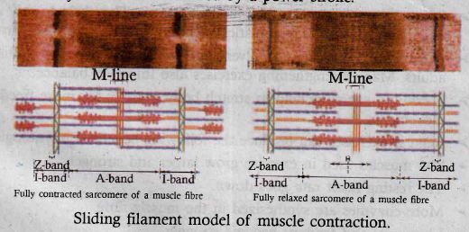
(xv) The power stroke (cross-bridge tilting) begins after the myosin head and hinge region tilt from a 90° angle to a 45° angle. The cross-bridge transforms into strong, high-force bond which allows the myosin head to swivel. When the myosin head swivels it pulls the attached actin filament towards the centre of the A-band.
(xvi) The myosin returns back to its relaxed state and releases ADP and phosphate ion. A new ATP molecule then binds to the head of the myosin and the cross-bridge is broken.
(xvii) At the end of each power stroke, each myosin head detaches from actin, then swivels back and binds to a new actin molecule to start another contraction cycle. This movement is similar to the motion of an oar on a boat.
(xviii) At the end of each power stroke, each myosin head detaches from actin, then swivels back and binds to a new actin molecule to start another contraction cycle.
(xix) The power stroke repeats many times until a muscle fibre contracts. The myosin heads bind, push and release actin molecules over and over as the thin filaments move toward the centre of the sarcomere.
(xx) The repeated formation of cross-bridge cycles cause the sliding of the filaments only but there is no change in the lengths of either the thick or thin filaments.
(xxi) The Z-discs attached to the actin filaments are also pulled inwards from both the sides, causing the shortening of the sarcomere (i.e. contraction). This process continues as long as the muscle receives the stimuli and with a steady flow of calcium ions.
(xxii) When motor impulse stops, the calcium ions are pumped back into the sarcoplasmic reticulum, results in the masking of the active sites of the actin filaments. The myosin head fails to bind with the active sites of actin and these changes cause the return of Z-discs back to their original position, i.e. relaxation.
19.
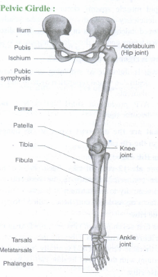
1. The pelvic girdle is a heavy structure specialized for weight bearing. It is composed of two hip bones called coxal bones that secure the lower limbs to the axial skeleton. Together, with the sacrum and coccyx, the hip bones form the basin-like bony pelvis.
2. Each coxal bone consists of three fused bones, ilium, ischium and pubis. At the point of fusion of ilium, ischium, and pubis a deep hemispherical socket called the acetabulum is present on the lateral surface of the pelvis.
3. It receives the head of the femur or thigh bone at the hip joint and helps in the articulation of the femur.
4. The ilium is the superior flaring portion of the hip bone. Each ilium forms a secure joint with the sacrum posteriorly. The ischium is a curved bar of bone. The V-shaped pubic bones articulate anteriorly at the pubic symphysis.
The Lower limb:
1. The lower limb consists of 30 bones which carries the entire weight of the erect body.
2. The bones of the lower limbs are thicker and stronger than the upper limbs. The three segments of each lower limb are the thigh, the leg or the shank and the foot.
3. The femur is the single bone of the thigh. It is the largest, longest and strongest bone in the body. The head of femur articulates with the acetabulum of the pelvis to form the hip joint.
4. Two parallel bones, the tibia and fibula, form the skeleton of the shank. A thick, triangular patella forms the kneecap, which protects the knee joint anteriorly and improves the leverage of thigh muscles acting across the knee.
5. The foot includes the bones of ankle, the tarsus, the metatarsus and the phalanges or toe bones. The foot supports our body weight and acts as a lever to propel the body forward while walking and running.
6. The tarsus is made up of seven bones called tarsals. The metatarsus consists of five bones called metatarsals. The arrangement of the metatarsals is parallel to each other. There are 14 phalanges in the toes which are smaller than those of the fingers.
20.
21.
1. Malfunctioning of the kidneys can lead to accumulation of urea and other toxic substances, leading to kidney failure. In such patients toxic urea can be removed from the blood by a process called haemodialysis
2. A dialysing machine or an artificial kidney is connected to the patient's body. A dialysing machine consists of a long cellulose tube surrounded by the dialysing fluid in a water bath.
3. The patient's blood is drawn from a convenient artery and pumped into the dialysing unit after adding an anticoagulant like heparin.
4. The tiny pores in the dialysis tube allows small molecules such as glucose, salts and urea to enter into the water bath, whereas blood cells and protein molecules do not enter these pores.
5. This stage is similar to the filtration process in the glomerulus. The dialysing liquid in the water bath consists of solution of salt and sugar in correct proportion in order to prevent loss of glucose and essential salts from the blood.
6. The cleared blood is then pumped back to the body through a vein.
22.
A. Aortic arch
B. Left Pulmonary Artery
C. Left Pulmonary Vein
D. Semilunar Valve
E. Left Ventricle
F. Right Ventricle
G. Inferior Vena Cava
23.
24.
(i) The process of elimination of nitrogenous wastes, salt and water balance from the body is called excretion.
(ii) This system consists of a paired kidneys, ureters, urinary bladder and cloaca.
(iii) Kidneys are dark red, long, flat organs situated on either sides of the vertebral column.
(iv) Kidneys are mesonephric type.
(v) The functional excretory units of kidneys are nephrons.
(vi) Nephrons separate nitrogenous wastes from the blood and excrete urea so frogs are ureotelic.
(vii) A paired long ureter emerges from the kidneys and opens into the cloaca.
(viii) A thin walled unpaired urinary bladder is present ventral to the rectum.
(ix) Rectum opens into the cloaca.
25.
1. The respiratory system of cockroach is well developed compared with other terrestrial insects.
2. Branched tubes known as trachea open through 10 pairs of small holes called spiracles or stigmata, present on the lateral side of the body.
3. Terminal branches of tracheal tubes are called tracheoles which carry oxygen to the entire body. The spiracles open and close by valves regulated by sphincter or spiracular muscles. Each tracheole is filled with a watery fluid through which exchange of gases takes place.
4. During high muscular activity, a part of the fluid is drawn into the tissues to enable more oxygen intake and rapid diffusion.
5. The passage of air in the tracheal system is:

26.
Classification of connective tissue and their functions Introduction:
(i) Connective tissue develops from the middle layer called mesoderm.
(ii) It is widely distributed in the body.
(iii) This tissue includes four classes.
(iv) The connective tissue includes fats and fibrous tissues
Four main classes are
i. Cartilage
ii. Bones
iii. Blood
iv. Other minor connective tissues
Components:
Main components are of three types, they are
1. Fibres
2. Ground substance or matrix
3. Cells
(i) Three fibres provide support in the matrix, they are collagen, elastic and reticular fibres.
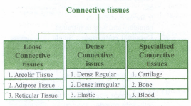
(ii) Main types of connective tissue are of three types
A) loose connective tissue
B) Dense connective tissue
C) Specialized connective tissue.
(A) Loose connective tissue:
(i) This tissue includes Areolar, Adipose and Reticular tissue.
(ii) Cells and fibres are loosely arranged in a semi fluid matrix.
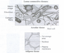
(i) Areolar tissue
(i) It acts as a support frame work for epithelium
(ii) It also acts as a reservoir of water and salts for the surrounding body tissues hence it is aptly called as tissue fluid.
(iii) It is a binding tissue.
(iv) Fibroblasts, macrophages and mast cells are its inclusions.
(ii) Adipose tissue
(i) It is found below the skin.
(ii) Cells are called as 'fat cells' or adipocytes.
(iii) 90% of this tissue mass is rich in fats.
(iv) This tissue stores fats which are unutilized nutrients for the metabolism of the body.
(v) This tissue is richly vascularised to exhibit high metabolic activity.
(vi) Under starvation this tissue provides fuel as energy rich substance.
(vii) It is found surrounding the kidneys, eyeball, heart, etc.
Divisions
(a) White fat or white adipose tissue: Cells contain less mitochondria, stores fat.
(b) Brown fat or Brown adipose tissue: Cells contain more mitochondria, oxidise fats to release heat energy (it is called as non-shivering thermogenesis)
(iii) Reticular:
(i) It forms an internal frame work (stroma) that supports the blood cells like lymphocytes in the lymph nodes, spleen and bone marrow.
(B) Dense connective tissue
(i) Fibres and fibroblasts are compactly packed
(ii) Divisible into i) Regular ii) Irregular iii) Elastic
i) Dense regular connective tissue:
(i) Regular type mainly contains collagen fibres in rows between parallel bundles of tissues.
(ii) It contains few elastic fibres.
(iii) Its major cell type is fibroblast which attaches to muscles.
(iv) It also withstands great tensile stress when pulling force is applied in one direction.
(v) It is found in tendon (bone and muscle are connected) and ligament (bone and bone are connected).
ii) Dense irregular connective tissue:
(i) Irregular type contains thick collagen fibres and the major cell type is fibroblast.
(ii) It is able to withstand tension exerted in many directions.
(iii) It provides structural strength.
(iv) Its elastic fibres are found in the leathery dermis
(v) It forms fibrous capsules of organs like kidneys, bones, cartilages, muscles, nerves and joints
iii) Elastic connective tissue:
(i) Elastic connective tissue consists of bulk of elastic fibres
(ii) It permits recoil of tissues following stretching.
(iii) It maintains the pulsatile flow of blood through the arteries and the passive recoil of lungs following inspiration.
(iv) It is found in the walls of large arteries, ligaments associated with vertebral column and within the walls of the bronchial tubes.
(C) Specialised connective tissues
(i) Classified into 1) Cartilage 2) Bones 3) Blood
(i) Cartilage:
(i) It is solid and found as inter cellular material and it is pliable.
(ii) It resists compression.
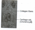
(iii) Cells are chondrocytes found in the small cavities of matrix.
(iv) Chondrocytes secrete matrix.
(v) Found in vertebrate embryos and then it is replaced by bones in adults.
(vi) It is found in the tip of nose, outer ear joints, external ear pinna, between adjacent bones of vertebral column, limbs and hands in adults.
(ii) Bones:
(i) A hard and non-pliable matrix rich in calcium salts and collagen fibres.
(ii) Calcium salts strengthen the bones and teeth.
(iii) Collagen fibres give mechanical strength.
(iv) It mainly provides structural frame to the body
(v) Bones support and protect the soft tissues of visceral organs.
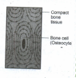
(vi) Osteocytes are bone cells found in the spaces called lacunae.
(vii) Bear the weight of the long bones of legs.
(viii) Bones interact with the skeletal muscles to bring about movements.
(ix) Bone marrow is the site of production of blood cells.
(iii) Blood:
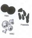
(i) It is a fluid connective tissue found with plasma, red blood cells, white blood cells and platelets.
(ii) It is a transportary medium of cardio vascular system.
(iii) It carries nutrients, wastes respiratory gases like O2 and CO2 throughout the body.
27.
Salient features of phylum Annelida:
1. Annelids were the first segmented animals to evolve. They are aquatic or terrestrial, free-living but some are parasitic.
2. They are triploblastic, bilaterally symmetrical, schizocoelomates and exhibit organ system level of body organisation. The coelom with coelomic fluid creates a hydrostatic skeleton and aids in locomotion.
3. Their elongated body is metamerically segmented and the body surface is divided into segment or metameres.
4. Internally the segments are divided from one another by partitions called septa. This phenomenon is known as metamerism. The longitudinal and circular muscles in the body wall help in locomotion.
5. Aquatic annelids like Nereis have lateral appendages called parapodia, which help in swimming. Chitinous setae in Earthworms, and suckers in Leech help in locomotion.
6. The circulatory system is of closed type and the respiratory pigments are hemoglobin and chlorocruorin.
7. Nervous system consists of paired ganglion connected by the lateral nerves to the double ventral nerve cord.
8. They reproduce sexually. Development is direct or indirect and includes a trochophore larva. Some are monoecious (earthworms) while some are dioecious (Nereis and Leech).
9. Eg: Lampito mauritii (earthworm), Nereis (sand worm), Hirudinaria (leech).
28.
(i) The word biology itself is a greek word where bios means life and logos means study.
(ii) The role for latin and greek words are used in classification especially in Binomial classification.
(iii) Named to understand and remember the scientific names of organisms.
(iv) The Binomial nomenclature (L.Bio-two; Nomen-Name) Vector animals and Plants is largely derived from latin and Greek words.
(v) Binomial nomenclature was originally codified in the works of Carolus Linnaeus (1753), Species Plantarum.
(vi) Latin is now used by classical scholars, certain purpose in botany and in the medicine field.
(vii) The purpose is to give every species a distinct name which will be recognized everywhere became common names abundant species vary from country to country or even from region to region within a country.
(viii) using latin and greek forms simply refresh the main language of science when this system was invented.
(ix) Other languages words now used are given grammatical endings derived from the classical languages. Ex: Blakea atton boroaghi.
29.
A multiple or composite fruit develops from the whole inflorescence along with its peduncle on which they are borne.
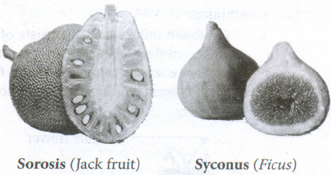
a) Sorosis: A fleshy multiple fruit which develops from a spike or spadix. The flowers fused together by their succulent perianth and at the same time, the axis bearing them become fleshy or juicy and the whole inflorescence forms a compact mass. Eg: Pineapple, Jackfruit, Mulberry
b) Syconus: A multiple fruit which develops from hypanthodium inflorescence. The receptacle develops further and converts into fleshy fruit which encloses a number of true fruit or achenes which develops from female flower of hypanthodium inflorescence. Eg: Ficus
30.
DNA barcoding is a taxonomic method that uses a very short genetic sequence from a standard part of a genome. The genetic sequence used to identify a plant is known as "DNA tags" or "DNA barcodes". Paul Hebert in 2003 proposed 'DNA barcoding' and he is considered as 'Father of barcoding'.
The gene region that is being used as an effective barcode in plants is present in two genes of the chloroplast, matK and rbcL, and have been approved as the barcode regions for plants.
Sequence of unknown species can be matched from submitted sequence in GenBank using BLAST (web programme for searching the closely related sequence).
Significance of DNA barcoding:
1. DNA barcoding greatly helps in identification and classification of organism.
2. It aids in mapping the extent of biodiversity.
DNA barcoding techniques require a large database of sequences for comparison and prior knowledge of the barcoding region.
However, DNA barcoding is a helpful tool to determine the authenticity of botanical material in whole, cut or powdered form.
31.
Structure of Mitochondria
1. It was first observed by A. Kolliker (1880), 1897, 1898 and named as mitochondria.
2. They are ovoid, rounded, rod-shaped and pleomorphic structures.
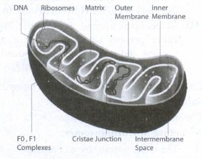
3. Mitochondrion consists of double membrane, the outer and inner membrane. The outer membrane is smooth, highly permeable to small molecules and it contains proteins called Porins, which form channels that allow free diffusion of molecules smaller than about 1000 daltons.
4. The inner membrane divides the mitochondrion into two compartments, outer chamber between two membranes and the inner chamber filled with matrix.
5. The inner membrane is convoluted (infoldings), called crista (plural: cristae). Cristae contain most of the enzymes for electron transport system.
6. Inner chamber of the mitochondrion is filled with proteinaceous material called mitochondrial matrix.
7. The inner membrane consists of stalked particles called elementary particles or Fernandez Moran particles, FI particles or Oxysomes. Each particle consists of a base, stem and a round head.
8. In the head ATP synthase is present for oxidative phosphorylation.
9. Inner membrane is impermeable to most ions, small molecules and maintains the proton gradient that drives oxidative phosphorylation Mitochondria contain 73% of proteins, 25-30% of lipids, 5-7% of RNA, DNA (in traces) and enzymes (about 60 types).
10. Mitochondria are called powerhouse of a cell, as they produce energy-rich ATP. All the enzymes of Kreb's cycle are found in the matrix except succinate dehydrogenase.
11. Mitochondria consist of circular DNA and 70S ribosome. They multiply by fission and replicate by strand displacement model. Because of the presence of DNA it is semi-autonomous organelle.
32.
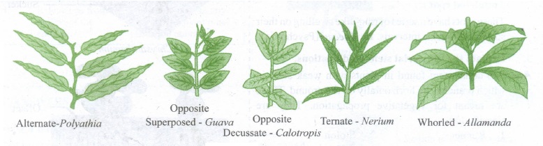
1. The arrangement of leaves on the stem or the branches is known as phyllotaxy. (Gk. Phyllon = leaf, taxis = arrangement).
2. Phyllotaxy is to avoid overcrowding of leaves so as to expose the leaves to maximum sunlight for photosynthesis.
3. The four main types of phyllotaxy are Alternate, Opposite, Ternate, Whorled.
Alternate phyllotaxy:
In this type there is only one leaf per node and leaves on the successive nodes are arranged alternately to each other. Spiral arrangement of leaves show vertical rows are called orthostichies. They are two types.
(a) Alternate spiral: In which the leaves are arranged alternately in a spiral manner. Eg: Hibiscus.
(b) Alternate distichous or Bifarious: In which the leaves are organized alternatively in two rows on either side of the stem. Eg: Polyalthia.
Opposite phyllotaxy: In this type each node possess two leaves opposite to each other. They are organized in two different types.
(a) Opposite superposed: The pair of leaves arranged in succession are in the same direction, that is two opposite leaves at a node lie exactly above those at the lower node. Eg: Psidium.
(b) Opposite decussate: In this type of phyllotaxy one pair of leaves is placed at right angles to the next upper or lower pair of leaves. Eg: Calotropis.
Ternate phyllotaxy: In this type there are three leaves attached at each node. Eg: Nerium.
Whorled (verticillate) type of phyllotaxy: In this type more than three leaves are present in a whorl at each node forming a circle or whorl. Eg: Allamanda.
33.
Mycorrhizae:
1. The Symbiotic association between fungal mycelium and roots of plants is called as mycorrhizae.
2. In this relationship fungi absorb nutrition from the root and in turn the hyphal network of the fungi helps the plant to absorb water and mineral nutrients from the soil. Mycorrhizae are classified into three types.
They are,
1. Ectomycorrhizae,
2. Endomycorrhizae,
3. Ectendomycorrhizae.
1. Ectomycorrhizae:
The fungal mycelium forms a dense sheath around the root called mantle. The hypha network penetrate the intercellular spaces of the epidermis and cortex to form Hartig net. Eg: Pisolithus tinctorius.
2. Endomycorrhizae:
the hyphae grow mainly inside the roots, penetrate the outer cortical cells of the plant root. A small portion of the mycelium is found outside the root. This form is also called Vesicular Arbuscular Mycorrhizal fungi (VAM Fungi) due to the presence of Vesicle or arbuscule like haustoria.
(i) Arbuscular mycorrhizae (VAM) Eg: Gigaspora
(ii) Ericoid mycorrhizae- Eg: Oidiodendron
(iii) Orchid mycorrhizae - Eg: Rhizoctonia
3. Ectendomycorrhizae:
The fungi form both mantle and also penetrates the cortical cells.
34.
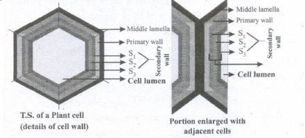
Cell wall is the outermost protective cover of cell. It was first observed by Robert Hooke. In plants it is made up of cellulose, hemicellulose, pectin, lignin, cutin, suberin and silica. In a plant cell wall shows three distinct regions (a) Primary wall (b) Secondary wall (c) Middle lamellae
(a) Primary wall:
(i) It is the first layer inner to middle lamellae, primarily consisting of loose network of cellulose microfibrils in a gel matrix.
(ii) It is thin, elastic and extensible. In most plants the microfibrils are made up of cellulose oriented differently based on shape and thickness of the wall.
(iii) The matrix of the primary wall is composed of hemicellulose, pectin, glycoprotein and water.
(iv) Hemicellulose binds the microfibrils with matrix and glycoproteins control the orientation of microfibrils while pectin serves as filling material of the matrix.
(v) Cells such as parenchyma and meristems have only primary wall.
Plant Cell Wall
b. Secondary wall :
(i) Secondary wall is laid during maturation. It plays a key role in determining the shape of a cell. It is thick, inelastic and is made up of cellulose and lignin.
(ii) The secondary wall is divided into three sub layers termed as SI' S2 and S3 where the cellulose microfibrils are compactly arranged with different orientation forming a laminated structure and the cell wall strength is increased.
c. Middle lamellae:
(i) It is the outermost layer made up of calcium and magnesium pectate, deposited at the time of cytokinesis.
(ii) It is a thin amorphous layer which cements two adjacent cells. It is optically inactive (isotropic).
(iii) Plasmodesmata and Pits: Plasmodesmata act as a channel between the protoplasm of adjacent cells through which many substances pass through.
(iv) Moreover, at few regions the secondary wall layer is laid unevenly whereas the primary wall and middle lamellae are laid continuously such regions are called pits
(v) The pits of adjacent cells are opposite to each other. Each pit has a pit chamber and a pit membrane.
(vi) The pit membrane has many minute pores and thus they are permeable. The pits are of two types namely simple and bordered pit.
35.
Tobacco mosaic virus was discovered in 1892 by Dmitri Ivanovsky from Tobacco plant.
Structure of Tobacco mosaic virus:
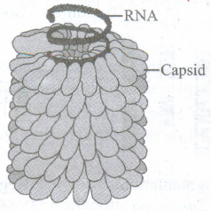
1. TMV is a rod-shaped helical virus measuring about 280 x 150\(\mu\)m with a molecular weight of 39 x 106 Daltons.
2. The virion is made up of two constituents, a protein coat called capsid and a core called nucleic acid.
3. The capsid is made up of approximately 2130 identical protein subunits called capsomeres.
4. The Nucleic acid consists of central single stranded RNA molecule.
5. The genetic information necessary for the formation of a complete TMV particle is contained in its RNA. It has 6,500 nucleotides.
36.
| S.No | Plants | Products | Uses |
| 1. | Cycas circinalis, Cycas revoluta | Sago | Starch used as food |
| 2. | Pinus gerardiana | Roasted seed | Used as a food |
| 3. | Abies balsamea | Resin | Used as mounting medium in permanent slide |
| 4. | Pinus insularis, Pinus roxburghii | Rosin and Turpentine | Paper sizing and varnishes |
| 5. | Araucaria, Picea | Tannins | Bark yield tannins and is used in Leather industries |
| 6. | Taxus brevifolia | Taxol | Drug used for cancer treatment |
| 7. | Ephedra gerardiana | Ephedrine | For the treatment of asthma, bronchitis |
| 8. | Pinus roxburghii | Oleoresin | Used to make soap, varnishes and printing ink |
| 9. | Pinus roxburghii, Picea smithiana | Wood pulp | Used to make papers |
| 10. | Cedrus deodara | Wood | Used to make doors, boats and railway sleepers |
| 11. | Cedrus atlantica | Oil | Used in perfumery |
| 12. | Thuja, Cupressus | Decorative | Ornamental plants |
37.
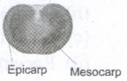
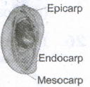
The fruits are derived from single pistil where the pericarp is fleshy, succulent and differentiated into epicarp, mesocarp and endocarp. It is subdivided into the following.
(i) Berry: Fruit develops from bicarpellary or multicarpellary, syncarpous ovary. Here the epicarp is thin, the mesocarp and endocarp remain undifferentiated. They form a pulp in which the seeds are embedded. Eg: Tomato, Grapes, Brinjal.
(ii) Drupe: Fruit develops from monocarpellary, superior ovary. It is usually one seeded. Pericarp is differentiated into outer skinny epicarp, fleshy and pulpy mesocarp and hard and stony endocarp around the seed. Eg: Mango, Coconut.
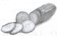
(iii) Pepo: Fruit develops from tricarpellary inferior ovary. Pericarp turns leathery or woody which encloses, fleshy mesocarp and smooth endocarp. Eg: Cucumber, Watermelon, Bottle gourd, Pumpkin.
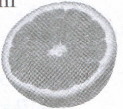
(iv) Hesperidium:Fruit develops from multicarpellary, multilocular, syncarpous, superior ovary. The fruit wall is differentiated into leathery epicarp with oil glands, a middle fibrous mesocarp. The endocarp forms distinct chambers, containing juicy hairs. Eg: Orange, Lemon.
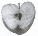
(v) Pome: It develops from multicarpellary, syncarpous, inferior ovary. The receptacle also develops along with the ovary and becomes fleshy, enclosing the true fruit. In pome the epicarp is thin skin like and endocarp is cartilaginous. Eg: Apple, Pear.
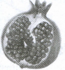
(vi) Balausta: A fleshy indehiscent fruit developing from multicarpellary, multilocular inferior ovary whose pericarp is tough and leathery. Seeds are attached irregularly with testa being the edible portion. Eg: Pomegranate.
38.
Features of DNA:
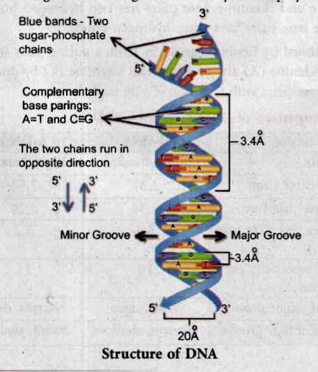
1. If one strand runs in the 5'- 3' direction, the other runs in 3'- 5' direction and thus are antiparallel (they run in opposite direction). The 5' end has the phosphate group and 3' end has the OH group.
2. The angle at which the two sugars protrude from the base pairs is about 120°, for the narrow angle and 240° for the wide angle. The narrow-angle between the sugars generates a minor groove and the large angle on the other edge generates major groove.
3. Each base is 0.34 nm apart and a complete turn of the helix comprises 3.4 nm or 10 base pairs per turn in the predominant B form of DNA.
4. DNA helical structure has a diameter of 20 A° and a pitch of about 34 A°. X-ray crystal study of DNA takes a stack of about 10 bp to go completely around the helix (360°).
5. Thermodynamic stability of the helix and specificity of base pairing includes
(i) the hydrogen bonds between the complementary bases of the double helix
(ii) stacking interaction between bases tend to stack about each other perpendicular to the direction of helical axis. Electron cloud interactions ((Π – Π)) between the bases in the helical stacks contribute to the stability of the double helix.
6. The phosphodiester linkages give an inherent polarity to the DNA helix. They form strong covalent bonds, give the strength and stability to the polynucleotide chain.
7. Plectonemic coiling - The two strands of the DNA are wrapped around each other in a helix, making it impossible to simply move them apart without breaking the entire structure. Whereas in paranemic coiling the two strands simply lie alongside one another, making them easier to pull apart.
8. Based on the helix and the distance between each turn the DNA is of three forms - A DNA, B DNA and Z DNA.
39.
(i) The symbiotic association between algae and fungi is called lichens.
(ii) The algal partner is called Phycobiont or Photobiont and the fungal partner is called Mycobiont.
(iii) Algae provide nutrition for fungal partner in turn fungi provide protection and also help to fix the thallus to the substratum through rhizines.
(iv) Asexual reproduction takes place through fragmentation, soredia and Isidia. Phycobionts reproduce by akinetes, hormogonia, aplanospore, etc., Mycobionts undergo sexual reproduction and produce ascocarps.
Classification:
(i) Based on the habitat lichens are classified into following types: Corticolous (on Bark) Lignicolous (on Wood) Saxicolous (on rocks) Terricolous (on ground) Marine (on siliceous rocks of sea) Fresh water (on siliceous rock of fresh water).
(ii) On the basis of morphology of the thallus they are divided into Leprose (a distinct fungal layer is absent) Crustose-crust like; Foliose-leaf like; Fruticose-branched pendulous shrub like.
(iii) The distribution of algal cells distinguishes lichens into two forms namely Homoiomerous (Algal cells evenly distributed in the thallus) and Heteromerous (a distinct layer of algae and fungi present)
(iv) If the fungal partner of lichen belongs to ascomycetes, it is called Ascolichen and if it is basidiomycetes it is called Basidiolichen.
40.
Factors affecting the rate of enzyme reactions: Enzymes are sensitive to environmental conditions. It could be affected by temperature, pH, substrate concentration and enzyme concentration. The rate of enzyme reaction is measured by the amount of substrate changed or amount of product formed during a period of time.
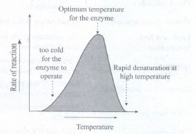
1. Temperature: Heating increases molecular motion. Thus the molecules of the substrate and enzyme move more quickly resulting in a greater probability of a reaction occurring. The temperature that promotes maximum activity is referred to as optimum temperature.
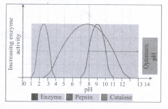
2. pH: The optimum pH is that at which the maximum rate of reaction occurs. Thus the pH change leads to an alteration of enzyme shape, including the active site. If extremes of pH are encountered by an enzyme, then it will be denatured.
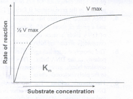
3. Substrate concentration: For a given enzyme concentration, the rate of an enzyme reaction increases with increasing substrate concentration.
4. Enzyme concentration: The rate of reaction is directly proportional to the enzyme concentration.
5. Michaelis-Menten Concept: When the initial rate of reaction of an enzyme is measured over a range of substrate concentrations (with a fixed amount of enzyme) and the results plotted on a graph. With increasing substrate concentration, the velocity increases - rapidly at lower substrate concentration.
6. Inhibitors of Enzyme: Certain substances present in the cells may react with the enzyme and lower the rate of reaction. These substances are called inhibitors. It is of two types competitive and non-competitive.
41.
Habit : Twining climber
Root : Branched tap root system having nodules.
Stem : Aerial, weak stem and a twiner
Leaf : Imparipinnately compound, alternate, stipulate showing reticulate venation. Leaflets are stipellate. Petiolate and stipels are pulvinated.
Inflorescence : Solitary and axillary
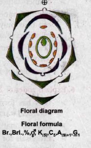
Flower : Bracteate, bracteolate, bracteoles usually large, pedicellate, heterochlamydeous, complete, bisexual, pentamerous, zygomorphic and hypogynous.
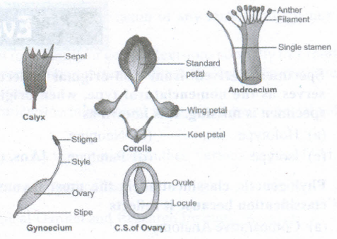
Calyx : Sepals 5, synsepalous, green showing valvate aestivation. Odd sepal is anterior in position.
Corolla : Petals 5, white or blue polypetalous, irregular papilionaceous corolla showing descendingly imbricate aestivation.
Androecium : Stamens 10, diadelphous (9)+1 nine stamens fused to form a bundle and the tenth stamen is free. Anthers are dithecous, basifixed, introrse and dehiscing by longitudinal slits.
Gynoecium : Monocarpellary, unilocular, with many ovules on marginal placentation, ovary superior, style simple and incurved with feathery stigma.
Fruit : Legume
Seed : Non-endospermous, reniform.
42.
43.
(i) Pachytene:
1. At this stage bivalent chromosomes are clearly visible as tetrads. Bivalent of meiosis I consists of 4 chromatids and 2 centromeres.
2. Synapsis is completed and recombination nodules appear at a site where crossing over takes place between non-sister chromatids of homologous chromosomes.
3. Recombination of homologous chromosomes is completed by the end of the stage but the chromosomes are linked at the sites of crossing over. This is mediated by the enzyme recombinase.
(ii) Diplotene:
1. Synaptonemal complex disassembles and dissolves. The homologous chromosomes remain attached at one or more points where crossing over has taken place.
2. These points of attachment where 'X' shaped structures occur at the sites of crossing over is called Chiasmata. Chiasmata are chromatin structures at sites where recombination has taken place.
44.
Occurs in distal convoluted tubule of nephron potassium is excreted and sodium is absorbed.
45.
(i) Nephrons of kidneys absorb and reabsorb blood plasma (55%) and handover it to venules of renal vein so that an average blood volume is maintained.
(ii) Oxygenated blood flows in the arterial system expresses the systolic pressure (120mm Hg) to balance the normal blood pressure towards the body organs.
46.
| Zoo | Wild life sanctuary |
| Wild animals kept in protected environments under human care Eg: Zoological parks | Wild animals are maintained in the protected areas under various projects for various animals and maintained by the services of Government. |
47.
48.
| Stages | ATP | NADH2 | FADH2 | Total ATPs |
| Glycolysis Link reaction Kreb's cycle |
2 - 2 |
2 \(\times\) 2.5 2 \(\times\) 2.5 6 \(\times\) 2.5 |
0 |
7 5 20 |
| 4 ATPs | 25 ATPs | 3 ATPs | 32 ATPs |
49.
Economic importance of Silk:
1. Silk fibers are utilized in preparing silk clothes. Silk fibers are now combined with other natural or synthetic fibers to manufacture clothes like Teri-silk, Cot-silk etc.
2. Silk is dyed and printed to prepare ornamented fabrics. They are generally made from Eri-silk or spun silk.
2. Silk is used in industries and for military purposes.
3. It is used in the manufacture of fishing fibers, parachutes, cartridge bags, insulation coils for telephone, wireless receivers, tyres of racing cars, filter fibres, in medical dressings and as suture materials.
50.
(i) Eggs or young ones produced by an oviparous (egg laying)and viviparous (directly yield young ones) female animals may not be equal.
(ii) Numerous eggs are laid by oviparous, so the number of produced offsprings is indefinite.
(iii) Eggs laid by oviparous, female may not develop into adults due to predators.
(iv) Oviparous female includes polyembryony.
(v) Viviparous produced by females need protection from predators associated with parental cares.
(vi) Development of young ones of oviparous female occurs in environment whereas the development of young ones of viviparous female occurs in the female reproductive system of adults.

11th Standard Syllabus & Materials
11th Standard
TN 11th Tamil பீடு பெற நில் - செய்யுள் - காவடிச்சிந்து Important Questions And Answers Study Material - QB365 Set A
NEW11th Standard
TN 11th Tamil பீடு பெற நில் - உரைநடை - மலை இடப்பெயர்கள் : ஓர் ஆய்வு Important Questions And Answers Study Material - QB365 Set A
NEW11th Standard
TN 11th Tamil மாமழை போற்றுதும் - துணைப்பாடம் - யானை டாக்டர் Important Questions And Answers Study Material - QB365 Set A
NEW11th Standard
TN 11th Tamil மாமழை போற்றுதும் - செய்யுள் - ஐங்குறுநூறு Important Questions And Answers Study Material - QB365 Set A



Tamilnadu Stateboard 11th Standard Subjects

Maths

Commerce

Economics
Biology

Business Maths and Statistics

Accountancy

Computer Science

Physics

Chemistry
Maths
Biology
Economics
Physics
Chemistry

History
Business Maths and Statistics

Computer Science
Accountancy

Computer Applications
History
Computer Technology
Commerce
Computer Applications
Computer Technology
Tamil

English

French
Tamilnadu Stateboard Standards