 100% FREE ACCESS
11th Standard Syllabus & Materials
11th Standard
TN 11th Tamil இயற்கை வேளாண்மை,சுற்றுச்சூழல் -செய்யுள் - மனோன்மணீயம் Important Questions And Answers Study Material - QB365 Set A
NEW11th Standard
TN 11th Tamil என்னுயிர் என்பேன் -துணைப்பாடம் - இசைத்தமிழர் இருவர் Important Questions And Answers Study Material - QB365 Set A
NEW11th Standard
TN 11th Tamil மொழி கலை -செய்யுள் - ஒவ்வொரு புல்லையும் Important Questions And Answers Study Material - QB365 Set A
NEW11th Standard
TN 11th Tamil பீடு பெற நில் - இலக்கணம் - பகுபத உறுப்புகள் Important Questions And Answers Study Material - QB365 Set A
NEW11th Standard
TN 11th Tamil பீடு பெற நில் - துணைப்பாடம் - வாடிவாசல் Important Questions And Answers Study Material - QB365 Set A
NEW11th Standard
TN 11th Tamil பீடு பெற நில் - செய்யுள் - குறுந்தொகை Important Questions And Answers Study Material - QB365 Set A

Published on: 13/05/2022
QB365 provides detailed and simple solution for every Creative Questions in class 11 Biology Subject. It will helps to get more idea about question pattern in every Creative questions with solution.
latest Creative QuestionsDownload Tamil Nadu 11th Standard Biology question papers, model tests, one-mark questions, important questions, and public exam papers in PDF format. Free study materials and answer keys for TN State Board students.
Questions + Answers key
Take MCQ Biology Test1.
How the nervous and endocrine system involve in the human cardiac function?
2.
Explain the process of coagulation of blood.
3.
Explain the recording of electrocardiogram.
4.
Explain about cardiac output (CO).
5.
Explain Erythroblastosis foetalis.
1.
1. The type of heart in human is myogenic because the heart beat originates from the muscles of the heart.
2. The nervous and endocrine systems work together with paracrine signals (metabolic activity) to influence the diameter of the arterioles and alter the blood flow.
3. The neuronal control is achieved through autonomic nervous system (sympathetic and parasympathetic). Sympathetic neurons release nor-epinephrine and adrenal medulla releases epinephrine.
4. The two hormones bind to β - adrenergic receptors and increase the heart rate.
5. The parasympathetic neurons secrete acetylcholine that binds to muscarinic receptors and decreases the heart beat.
6. Vasopressin and angiotensin II, involved in the regulation of the kidneys, results in vasoconstriction while natriuretic peptide promotes vasodilation.
7. Vagus nerve is a parasympathetic nerve that supplies the atrium especially the SA and the AVnodes.
2.
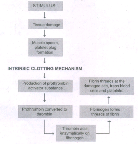
If you cut your finger or when you get yourself hurt, your wound bleeds for some time after which it stops to bleed. This is because the blood clots or coagulates in response to trauma. The mechanism by which excessive blood loss is prevented by the formation of clot is called blood coagulation or clotting of blood. The clotting process begins when the endothelium of the blood vessel is damaged and the connective tissue in its wall is exposed to the blood. Platelets adhere to collagen fibres in the connective tissue and release sub stances that form the platelet plug which provides emergency protection against blood loss. Clotting factors released from the clumped platelets or damaged cells mix with clotting factors in the plasma. The protein called prothrombin is converted to its active form called thrombin in thepresence of calcium and vitamin K. Thrombin helps in the conversion of fibrinogen to fibrin threads. The threads of fibrins become interlinked into a patch that traps blood cell and seals the injured vessel until the wound is healed. After sometime fibrin fibrils contract, squeezing out a straw-coloured fluid through a meshwork called serum (Plasma without fibrinogen is called serum). Heparin is an anticoagulant produced in small quantities by mast cells of connective tissue which prevents coagulation in small blood vessels.
3.
An electrocardiogram (ECG) records the electrical activity of the heart over a period of time using electrodes placed on the skin, arms, legs, and chest. It records the changes in electrical potential across the heart during one cardiac cycle. The special flap of muscle which initiates the heart beat is called as sinu-auricular node or SA node in the right atrium. It spreads as a wave of contraction in the heart. The waves of the ECG are due to depolarization and not due to contraction of the heart. This wave of depolarisation occurs before the beginning of contraction of the cardiac muscle. A normal ECG shows 3 waves designated as P wave, QRS complex and T wave.
P Wave (atrial depolarisation): It is a small upward wave and indicates the depolarisation of the atria. This is the time taken for the excitation to spread through atria from SA node. Contraction of both atria lasts for around 0.8-1.0 sec.
PQ Interval (AV node delay): It is the onset of P wave to the onset of QRS complex. This is from the start of depolarisation of the atria to the beginning of ventricular depolarisation. It is the time taken for the impulse to travel from the atria to the ventricles (0.12-0.21sec). It is the measure of AV conduction time.
QRS Complex (ventricular depolarisation): No separate wave for atrial depolarisation in the ECG is visible. Atrial depolarisation occurs simultaneously with the ventricular depolarisation. The normal QRS complex lasts for 0.06-0.09 sec. QRS complex is shorter than the P wave because depolarisation spreads through the Purkinjie fibres. Prolonged QRS wave indicates delayed conduction through the ventricle, often caused due to ventricular hypertrophy or due to a block in the branches of the bundle of His.
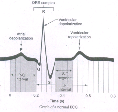
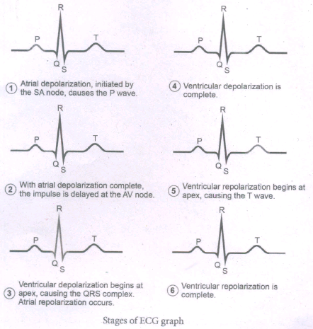
ST Segment: It lies between the QRS complex and T wave. It is the time during which all regions of the ventricles are completely depolarised and reflects the long plateau phase before repolarisation. In the heart muscle, the prolonged depolarisation is due to retardation of K+ efflux and is responsible for the plateau. The ST segment lasts for 0.09 sec.
T wave (ventricular depolarisation): It represents ventricular depolarisation. The duration of the T wave is longer than QRS complex because repolarisation takes place simultaneously throughout the ventricular depolarisation.
4.
(i) Definition: The amount of blood pumped out by each ventricle per minute is called cardiac output (CO).
(ii) Heart rate (HR) and stroke volume (SV) are the products of CO.
(iii) HR or pulse is the number of beats per minute.
(iv) So pulse pressure = Systolic pressure - Diastolic pressure.
(v) SV is the volume of blood pumped out by one ventricle with each beat.
(vi) SV depends on ventricular systole/contraction.
(vii) CO = HR x SV
(viii) SV represents the difference between EDV (amount of blood that collects in a ventricle during diastole).
(ix) SV = EDV - ESV.
(x) Frank-Starling law of the heart, the critical factor controlling SV is the degree to which the cardiac myocytes are stretched just before they contract.
(xi) The most important factor stretching cardiac muscle is the amount of blood returning to the heart and distending its ventricles venous return.
(xii) SV performs the doubling of circulation during venous return during physical exercise.
(xiii) A balance between cardiac output and venous return is maintained by the pumping action of heart.
(xiv) As the heart serves as a double pump, each side can fail independently of the other.
(xv) If the left side of the heart fails. It results in pulmonary congestion and similarly the right side of the heart fails it results in the peripheral congestion.
(xvi) Frank-Starling effect protects the heart from abnormal increase in blood volume.
5.
1. Rh factor is a protein (D antigen) present on the surface of the red blood cells in majority (80%) of humans.
2. This protein is similar to the protein present in Rhesus monkey, hence the term Rh.
3. Individuals who carry the antigen D on the surface of the red blood cells are Rh" (Rh positive) and the individuals who do not carry antigen D, are Rh" (Rh negative).
4. Rh factor compatibility is also checked before blood transfusion. When a pregnant women is Rh" and the foetus is Rh" incompatibility (mismatch) is observed.
5. During the first pregnancy, the Rh antigens of: the foetus does not get exposed to the mother's blood as both their blood are separated by placenta.
6. However, small amount of the foetal antigen becomes exposed to the mother's blood during the birth of the first child.
7. The mother's blood starts to synthesize D antibodies. But during subsequent pregnancies, the Rh antibodies from the mother (Rh") enters the foetal circulation and destroys the foetal RBCs.
8. This becomes fatal to the foetus because the child suffers from anaemia and jaundice. This condition is called erythroblastosis foetalis. This condition can be avoided by administration of anti D antibodies (Rhocum) to the mother immediately after the first childbirth.

11th Standard Syllabus & Materials
11th Standard
TN 11th Tamil பீடு பெற நில் - செய்யுள் - காவடிச்சிந்து Important Questions And Answers Study Material - QB365 Set A
NEW11th Standard
TN 11th Tamil பீடு பெற நில் - உரைநடை - மலை இடப்பெயர்கள் : ஓர் ஆய்வு Important Questions And Answers Study Material - QB365 Set A
NEW11th Standard
TN 11th Tamil மாமழை போற்றுதும் - துணைப்பாடம் - யானை டாக்டர் Important Questions And Answers Study Material - QB365 Set A
NEW11th Standard
TN 11th Tamil மாமழை போற்றுதும் - செய்யுள் - ஐங்குறுநூறு Important Questions And Answers Study Material - QB365 Set A



Tamilnadu Stateboard 11th Standard Subjects

Maths

Commerce

Economics
Biology

Business Maths and Statistics

Accountancy

Computer Science

Physics

Chemistry
Maths
Biology
Economics
Physics
Chemistry

History
Business Maths and Statistics

Computer Science
Accountancy

Computer Applications
History
Computer Technology
Commerce
Computer Applications
Computer Technology

Tamil

English

French
Tamilnadu Stateboard Standards