 100% FREE ACCESS
11th Standard Syllabus & Materials
11th Standard
TN 11th Tamil இயற்கை வேளாண்மை,சுற்றுச்சூழல் -செய்யுள் - மனோன்மணீயம் Important Questions And Answers Study Material - QB365 Set A
NEW11th Standard
TN 11th Tamil என்னுயிர் என்பேன் -துணைப்பாடம் - இசைத்தமிழர் இருவர் Important Questions And Answers Study Material - QB365 Set A
NEW11th Standard
TN 11th Tamil மொழி கலை -செய்யுள் - ஒவ்வொரு புல்லையும் Important Questions And Answers Study Material - QB365 Set A
NEW11th Standard
TN 11th Tamil பீடு பெற நில் - இலக்கணம் - பகுபத உறுப்புகள் Important Questions And Answers Study Material - QB365 Set A
NEW11th Standard
TN 11th Tamil பீடு பெற நில் - துணைப்பாடம் - வாடிவாசல் Important Questions And Answers Study Material - QB365 Set A
NEW11th Standard
TN 11th Tamil பீடு பெற நில் - செய்யுள் - குறுந்தொகை Important Questions And Answers Study Material - QB365 Set A
Published on: 08/06/2021
QB365 provides detailed and simple solution for every book back questions in class 11 Biology subject.It will helps to get more idea about question pattern in every book back questions with solution.
Download Tamil Nadu 11th Standard Biology question papers, model tests, one-mark questions, important questions, and public exam papers in PDF format. Free study materials and answer keys for TN State Board students.
Questions + Answers key
Take MCQ Biology Test1.
What is tubular secretion? Name the substances secreted through the renal tubules.
2.
3.
What is the difference between a Zoo and Wild Life Sanctuary?
4.
Why elephants and other wild animals are entering into human living area?
5.
6.
Identify the biological term
Excretion, glomerulus, urinary bladder, glomerular filtrate, ureters, urine, Bowman’s capsule, urinary system, reabsorption, micturition, osmosis, proteins.
a. A liquid which gathers in the bladder.
b. Produced when blood is filtered in a Bowman’s capsule.
c. Temporary storage of urine.
d. A ball of intertwined capillaries.
e. Removal of unwanted substances from the body.
f. Each contains a glomerulus.
g. Carry urine from the kidneys to the bladder.
h. Scientific term for urination.
i. Regulation of water and dissolved substances in blood and tissue fluid.
j. Consists of the kidneys, ureters and bladder.
k. Removal of useful substances from glomerular filtrate.
l. What solute the blood contains that are not present in the glomerular filtrate?
7.
Draw a neat labelled diagram of the digestive system of frog.
8.
Observe the animal below and answer the following questions.
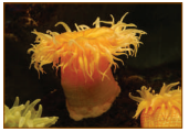
a. Identify the animal
b. What type of symmetry does this animal exhibit?
c. Is this animal Cephalized?
d. How many germ layers does this animal have?
e. How many openings does this animal's digestive system have?
f. Does this animal have neurons?
9.
Can we use recent molecular tools to identify and classify organisms?
10.
Explain the principle involved in PET scan
11.
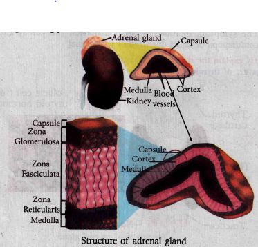
Name the layers of adrenal cortex and mention their secretions.
12.
Write the causes for diabetes mellitus and diabetes insipidus.
13.
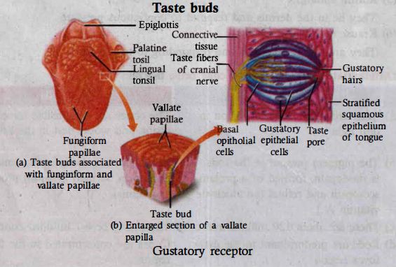
The sense of taste is considered to be the most pleasurable of all senses. Describe the structure of the receptor involved with a diagram.
14.
Explain the sliding-filament theory of muscle contraction.
15.
What are the bones that make the pelvic girdle?
16.
Discuss the various techniques adopted in cattle breeding.
17.
Animal husbandry is the science of rearing, feeding and caring, breeding and disease control of animals. It ensures supply of proper nutrition to our growing population through activities like increased production and improvement of animal products like milk, eggs, meat, honey, etc.
a. Poultry production depends upon the photoperiod. Discuss
b. Polyculture of fishes is of great importance. Discuss.
18.
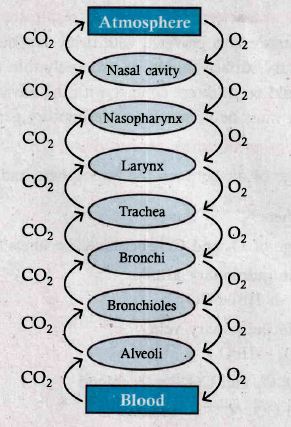
Sketch a flow chart to show the pathway of air flow during respiration.
19.
Arrange the following structures in the order that a drop of water entering the nephron would encounter them
e. Afferent arteriole
f. Bowman’s capsule
g. Collecting duct
h. Distal tubule
i. Glomerulus
j. Loop of Henle
k. Proximal tubule
l. Renal pelvis
20.
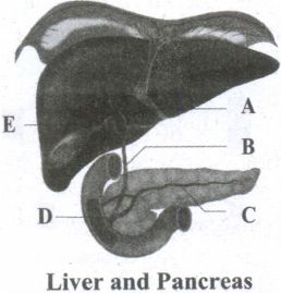
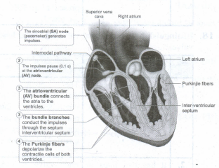
Label the given diagram.
21.
Select the correct biological term.
Cardiac muscle, atria, tricuspid systole, auricles, arteries, diastole, ventricles, bicuspid valve, pulmonary artery, cardiac cycle, semilunar valve, veins, pulmonary vein, capillaries, vena cava, aorta.
(a) The main artery of the blood
(b) Valves between the left atrium and ventricle
(c) Technical name for relaxation of the heart.
(d) Another name for atria.
(e) The main vein.
(f) Vessels which carry blood away from the heart
(g) Two names for the upper chambers of the heart
(h) Thick walled chambers of the heart.
(i) Carries blood from the heart to the lungs
(j) Takes about 0.8 sec to complete.
(k) Valves situated at the point where blood flows out of the heart.
(l) Vessels which carry blood towards the heart
(m) Carries blood from the lungs to the heart.
(n) The two lower chambers of the heart.
(o) Prevent blood from re-entering the ventricles after entering the aorta.
(p) Technical name for one heartbeat.
(q) Valves between right atrium and ventricles.
(r) Technical name for contraction of the heart.
(s) Very narrow blood vessels.
22.
23.
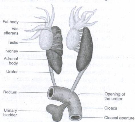
Explain the excretory system of frog.
24.
How respiration takes place in cockroach?
25.
Write the classification of connective tissue and their functions.
1.
Occurs in distal convoluted tubule of nephron potassium is excreted and sodium is absorbed.
2.
3.
| Zoo | Wild life sanctuary |
| Wild animals kept in protected environments under human care Eg: Zoological parks | Wild animals are maintained in the protected areas under various projects for various animals and maintained by the services of Government. |
4.
Lack of food and favourable climate, food competition, non availability of water, interference of affect of predators elephants and other wild animals enter into human living area.
5.
6.
(a) Urine
(b) Glomerular filtrate
(c) Urinary bladder
(d) afferent and efferent arterioles and venules of renal artery and renal vein.
(e) Excretion
(f) Bowman's capsule
(g) Ureters
(h) Micturition
(i) Balance of electrolytes minerals and organics
(j) excretory system
(k) micturition
(l) glucose, amino acid and liquids
7.
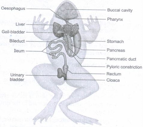
8.
a. Sea anemone
b. Radial symmetry
c. Not cephalized
d. Two
e. A single
f. Yes
9.
(i) Yes we can use recent molecular tools to identify and classify organisms.
(ii) Eg: Automated species identification tools electron microscopy (for cellular organelles and molecular structures by neo taxonomical tools) behaviour of organisms by ethology of taxonomical tools and development of electronic digital images and description by INOTAXA or e-Taxonomic resources.
10.
Positron Emission Tomographic Scanning (PET):
1. PET is also computerized imaging technique unlike CT.
2. Positron emission tomography (PET) is a nuclear medicine procedure based on the measurement of positron emission from radiolabelled tracer molecules.
11.
(i) A pair of adrenal glands are located at the anterior end of the kidneys. The outer region is the cortex and the inner region is the medulla.
(ii) The adrenal cortex has three distinct zones, they are 1. Zona glomerulosa 2. Zona fasciculata and 3. Zona reticularis.
(iii) Zona glomerulosa an outer thin layer constitutes about 15% of adrenal cortex and its secretes mineralocorticoids.
(iv) Zona fasciculata, the middle layer constitutes about 75% of adrenal cortex and secretes glucocorticoids such as cortisol, corticosterone and trace amounts of adrenal androgen and oestrogen.
(v) Zona Reticularis, an inner zone of adrenal cortex constitute about 10% of adrenal cortex and secretes the adrenal cortex androgen, trace amount of oestrogen and glucocorticoids.
12.
Diabetes mellitus:
1. It is otherwise known as hyperglycaemia.
2. It is caused due to lower secretion of insulin. By this process blood glucose level is elevated. There are two types of diabetes mellitus. Types I and Type II.
3. Type I diabetes is known Insulin dependent diabetes, caused by the lack of insulin secretion due to illness or some viral infections.
4. Type II diabetes is also known as Non-Insulin dependent diabetes, caused due to lower secretion of insulin.
5. The half-life period of insulin [in plasma] is only 6 minutes.
6. Symptoms of diabetes includes polyuria, polyphagia, polydipsia [excessive consumption of liquid due to thirst] ketosis [Breakdown of fat into glucose result in accumulation of ketone bodies in blood.]
Diabetes insipidus:
1. Diabetes insipidus is caused due to hypo secretion of vasopressin [ADH] from neurohypophysis
2. The symptom includes frequent urination [Polyuria] and excessive consumption of liquid due to thirst [polydipsia]
13.
1. The sense of taste is considered to be the most pleasurable of all senses.
2. The tongue is provided with many small projections called papillae which give the tongue an abrasive feel.
3. Taste buds are located mainly on the papillae which are scattered over the entire tongue surface.
4. Most taste buds are seen on the tongue few are scattered on the soft palate, inner surface of the cheeks, pharynx and epiglottis of the larynx.
5. Taste buds are flask-shaped and consist of 50 - 100 epithelial cells of two major types.
14.
Mechanism of muscle contraction
(i) Sliding filament theory in 1954, Andrew F. Huxley and Rolf Niedergerke proposed the sliding-filament theory to explain muscle contraction.
(ii) According to this theory, overlapping actin and myosin filaments of fixed length slide past one another in an energy requiring process, resulting in muscle contraction.
(iii) The contraction of muscle fibre is a remarkable process that helps in creating a force to move or to resist a load.
(iv) The force which is created by the contracting muscle is called muscle tension.
(v) The load is a weight or force that opposes contraction of a muscle.
(vi) Contraction is the creation of tension in the muscle which is an active process and relaxation is the release of tension created by contraction.
(vii) Muscle contraction is initiated by a nerve impulse sent by the central nervous system (CNS) through a motor neuron.
(viii) The junction between the motor neuron and the sarcolemma of the muscle fibre is called the neuromuscular junction or motor end plate.
(ix) When nerve impulse reaches a neuromuscular junction, acetylcholine is released.
(x) It initiates the opening of multiple gated channels in sarcolemma. The action potential travels along the T-tubules and triggers the release of calcium ions from the sarcoplasmic reticulum.
(xi) The released calcium ions bind to troponin on thin filaments. The tropomyosin uncovers the myosin-binding sites on thin filaments.
(xii) Now the active sites are exposed to the heads of myosin to form a cross-bridge. During cross-bridge formation actin and myosin form a protein complex called actomyosin.
(xiii) Utilizing the energy released from hydrolysis of ATP, the myosin head rotates until it forms a 90° angle with the long axis of the filament.
(xiv) In this position myosin binds to an actin and activates a contraction relaxation cycle which is followed by a power stroke.
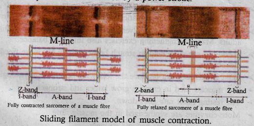
(xv) The power stroke (cross-bridge tilting) begins after the myosin head and hinge region tilt from a 90° angle to a 45° angle. The cross-bridge transforms into strong, high-force bond which allows the myosin head to swivel. When the myosin head swivels it pulls the attached actin filament towards the centre of the A-band.
(xvi) The myosin returns back to its relaxed state and releases ADP and phosphate ion. A new ATP molecule then binds to the head of the myosin and the cross-bridge is broken.
(xvii) At the end of each power stroke, each myosin head detaches from actin, then swivels back and binds to a new actin molecule to start another contraction cycle. This movement is similar to the motion of an oar on a boat.
(xviii) At the end of each power stroke, each myosin head detaches from actin, then swivels back and binds to a new actin molecule to start another contraction cycle.
(xix) The power stroke repeats many times until a muscle fibre contracts. The myosin heads bind, push and release actin molecules over and over as the thin filaments move toward the centre of the sarcomere.
(xx) The repeated formation of cross-bridge cycles cause the sliding of the filaments only but there is no change in the lengths of either the thick or thin filaments.
(xxi) The Z-discs attached to the actin filaments are also pulled inwards from both the sides, causing the shortening of the sarcomere (i.e. contraction). This process continues as long as the muscle receives the stimuli and with a steady flow of calcium ions.
(xxii) When motor impulse stops, the calcium ions are pumped back into the sarcoplasmic reticulum, results in the masking of the active sites of the actin filaments. The myosin head fails to bind with the active sites of actin and these changes cause the return of Z-discs back to their original position, i.e. relaxation.
15.
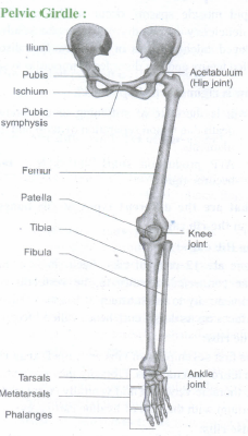
1. The pelvic girdle is a heavy structure specialized for weight bearing. It is composed of two hip bones called coxal bones that secure the lower limbs to the axial skeleton. Together, with the sacrum and coccyx, the hip bones form the basin-like bony pelvis.
2. Each coxal bone consists of three fused bones, ilium, ischium and pubis. At the point of fusion of ilium, ischium, and pubis a deep hemispherical socket called the acetabulum is present on the lateral surface of the pelvis.
3. It receives the head of the femur or thigh bone at the hip joint and helps in the articulation of the femur.
4. The ilium is the superior flaring portion of the hip bone. Each ilium forms a secure joint with the sacrum posteriorly. The ischium is a curved bar of bone. The V-shaped pubic bones articulate anteriorly at the pubic symphysis.
The Lower limb:
1. The lower limb consists of 30 bones which carries the entire weight of the erect body.
2. The bones of the lower limbs are thicker and stronger than the upper limbs. The three segments of each lower limb are the thigh, the leg or the shank and the foot.
3. The femur is the single bone of the thigh. It is the largest, longest and strongest bone in the body. The head of femur articulates with the acetabulum of the pelvis to form the hip joint.
4. Two parallel bones, the tibia and fibula, form the skeleton of the shank. A thick, triangular patella forms the kneecap, which protects the knee joint anteriorly and improves the leverage of thigh muscles acting across the knee.
5. The foot includes the bones of ankle, the tarsus, the metatarsus and the phalanges or toe bones. The foot supports our body weight and acts as a lever to propel the body forward while walking and running.
6. The tarsus is made up of seven bones called tarsals. The metatarsus consists of five bones called metatarsals. The arrangement of the metatarsals is parallel to each other. There are 14 phalanges in the toes which are smaller than those of the fingers.
16.
There are various techniques adopted in cattle breeding.
1. Inbreeding:
i. Breeding between animals of the same breed for 4-6 generations is called inbreeding. Inbreeding increases homozygosity and exposes the harmful recessive genes.
ii. Continuous inbreeding reduces fertility and even productivity, resulting in "inbreeding depression" .
iii. This can be avoided by breeding selected animals of the breeding population and they should be mated with superior animals of the same breed but unrelated to the breeding population. It helps to restore fertility and yield.
2. Outbreeding:
(i) The breeding between unrelated animals is called outbreeding.
(ii) Outbreeding helps to produce new and favourable traits, to produce hybrids with superior qualities and helps to create new breeds. In outbreeding, there are another steps also involved.
i. Outcrossing:
It is the breeding between unrelated animals of the same breed but having no common ancestry. The offspring of such a cross is called outcross. This method is suitable for breeding animals below average in productivity.
ii. Crossbreeding: Breeding between a superior male of one breed with a superior female of another breed. The crossbred progeny has superior traits.
iii. Interspecific hybridization: Method of breeding between male and female of two different species. For eg male donkey is crossed with female horse the offspring is called as mule.
3. Artificial insemination:
(i). Another breeding method is Artificial insemination is a technique in which the semen collected from the male is injected to the reproductive tract of the selected female.
1. It is economical low cost.
2. No semen are wasted.
3. Cross breeding and outbreeding are favourable in this method.
17.
(a) Photoperiod which means light plays a important functions in poultry farming.
1. Light influences the growth, development and production of chickens.
2. Light colour, duration and intensity play a role in poultry production.
3. It stimulates the internal cycle due to long day period.
4. Different coloured lights give the chickens to grow in proper way example blue colour gives them calming effect, blue-green keep them to grow and orange-red light stimulates them for their reproduction.
5. The photoperiod light also linked with temperature. The lamps light giving light with a cooling effect which generates a lot of heat in their body this impact on growth of chicks along with light colour and intensity of the light.
(b) Polyculture of fishes:
Polyculture is otherwise known as composite fish farming. Some selected fishes which are belonged to different species are stocked together in proper proportion in a fish pond. This mixed farming is termed as composite fish farming.
Importance:-
1. Polyculture utilized all available nutrients
2. No Competition among different species is found.
3. Catla catla, labeo rohita, and cirrhinus mrigala are the commonly used fish species for polyculture fish farming.
18.
19.
(e) Afferent arteriole
(i) Glomerulus
(f) Bowman's capsule
(k) Proximal tubule
(j) Loop of Henle
(h) Distal tubule
(g) Collecting duct
(l) Renal pelvis
20.
A. Common hepatic duct
B. Common bile duct
C. Pancreatic duct (duct of Wirsung)
D. Sphincter of Oddi
E. Cystic duct
21.
(a) Aorta
(b) Bicuspid
(c) Diastole
(d) Auricle
(e) Vena cava
(f) Arteries
(g) Atria /Auricles
(h) Ventricles
(i) Pulmonary Artery
(j) Cardiac cycle
(k) Semilunar Valves
(l) Veins
(m) Pulmonary vein
(n) Ventricles
(o) Semilunar Valve
(p) Cardiac cycle
(q) Tricuspid Valve
(r) Systole
(s) Capillaries
22.
23.
(i) The process of elimination of nitrogenous wastes, salt and water balance from the body is called excretion.
(ii) This system consists of a paired kidneys, ureters, urinary bladder and cloaca.
(iii) Kidneys are dark red, long, flat organs situated on either sides of the vertebral column.
(iv) Kidneys are mesonephric type.
(v) The functional excretory units of kidneys are nephrons.
(vi) Nephrons separate nitrogenous wastes from the blood and excrete urea so frogs are ureotelic.
(vii) A paired long ureter emerges from the kidneys and opens into the cloaca.
(viii) A thin walled unpaired urinary bladder is present ventral to the rectum.
(ix) Rectum opens into the cloaca.
24.
1. The respiratory system of cockroach is well developed compared with other terrestrial insects.
2. Branched tubes known as trachea open through 10 pairs of small holes called spiracles or stigmata, present on the lateral side of the body.
3. Terminal branches of tracheal tubes are called tracheoles which carry oxygen to the entire body. The spiracles open and close by valves regulated by sphincter or spiracular muscles. Each tracheole is filled with a watery fluid through which exchange of gases takes place.
4. During high muscular activity, a part of the fluid is drawn into the tissues to enable more oxygen intake and rapid diffusion.
5. The passage of air in the tracheal system is:

25.
Classification of connective tissue and their functions Introduction:
(i) Connective tissue develops from the middle layer called mesoderm.
(ii) It is widely distributed in the body.
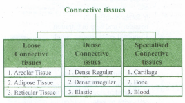
(iii) This tissue includes four classes.
(iv) The connective tissue includes fats and fibrous tissues
Four main classes are
i. Cartilage
ii. Bones
iii. Blood
iv. Other minor connective tissues
Components:
Main components are of three types, they are
1. Fibres
2. Ground substance or matrix
3. Cells
(i) Three fibres provide support in the matrix, they are collagen, elastic and reticular fibres.
(ii) Main types of connective tissue are of three types
A) loose connective tissue
B) Dense connective tissue
C) Specialized connective tissue.
(A) Loose connective tissue:
(i) This tissue includes Areolar, Adipose and Reticular tissue.
(ii) Cells and fibres are loosely arranged in a semifluid matrix.
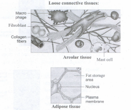
(i) Areolar tissue
(i) It acts as a support framework for epithelium
(ii) It also acts as a reservoir of water and salts for the surrounding body tissues hence it is aptly called as tissue fluid.
(iii) It is a binding tissue.
(iv) Fibroblasts, macrophages and mast cells are its inclusions.
(ii) Adipose tissue
(i) It is found below the skin.
(ii) Cells are called as 'fat cells' or adipocytes.
(iii) 90% of this tissue mass is rich in fats.
(iv) This tissue stores fats which are unutilized nutrients for the metabolism of the body.
(v) This tissue is richly vascularised to exhibit high metabolic activity.
(vi) Under starvation this tissue provides fuel as energy rich substance.
(vii) It is found surrounding the kidneys, eyeball, heart, etc.
Divisions
(a) White fat or white adipose tissue: Cells contain less mitochondria, stores fat.
(b) Brown fat or Brown adipose tissue: Cells contain more mitochondria, oxidise fats to release heat energy (it is called as non-shivering thermogenesis)
(iii) Reticular:
(i) It forms an internal framework (stroma) that supports the blood cells like lymphocytes in the lymph nodes, spleen and bone marrow.
(B) Dense connective tissue
(i) Fibres and fibroblasts are compactly packed
(ii) Divisible into i) Regular ii) Irregular iii) Elastic
i) Dense regular connective tissue:
(i) Regular type mainly contains collagen fibres in rows between parallel bundles of tissues.
(ii) It contains few elastic fibres.
(iii) Its major cell type is fibroblast which attaches to muscles.
(iv) It also withstands great tensile stress when pulling force is applied in one direction.
(v) It is found in tendon (bone and muscle are connected) and ligament (bone and bone are connected).
ii) Dense irregular connective tissue:
(i) Irregular type contains thick collagen fibres and the major cell type is fibroblast.
(ii) It is able to withstand tension exerted in many directions.
(iii) It provides structural strength.
(iv) Its elastic fibres are found in the leathery dermis
(v) It forms fibrous capsules of organs like kidneys, bones, cartilages, muscles, nerves and joints
iii) Elastic connective tissue:
(i) Elastic connective tissue consists of bulk of elastic fibres
(ii) It permits recoil of tissues following stretching.
(iii) It maintains the pulsatile flow of blood through the arteries and the passive recoil of lungs following inspiration.
(iv) It is found in the walls of large arteries, ligaments associated with vertebral column and within the walls of the bronchial tubes.
(C) Specialised connective tissues
(i) Classified into 1) Cartilage 2) Bones 3) Blood
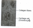
(i) Cartilage:
(i) It is solid and found as intercellular material and it is pliable.
(ii) It resists compression.
(iii) Cells are chondrocytes found in the small cavities of matrix.
(iv) Chondrocytes secrete matrix.
(v) Found in vertebrate embryos and then it is replaced by bones in adults.
(vi) It is found in the tip of nose, outer ear joints, external ear pinna, between adjacent bones of vertebral column, limbs and hands in adults.
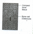
(ii) Bones:
(i) A hard and non-pliable matrix rich in calcium salts and collagen fibres.
(ii) Calcium salts strengthen the bones and teeth.
(iii) Collagen fibres give mechanical strength.
(iv) It mainly provides structural frame to the body
(v) Bones support and protect the soft tissues of visceral organs.
(vi) Osteocytes are bone cells found in the spaces called lacunae.
(vii) Bear the weight of the long bones of legs.
(viii) Bones interact with the skeletal muscles to bring about movements.
(ix) Bone marrow is the site of production of blood cells.
(iii) Blood:
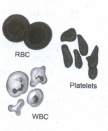
(i) It is a fluid connective tissue found with plasma, red blood cells, white blood cells and platelets.
(ii) It is a transport medium of cardiovascular system.
(iii) It carries nutrients, wastes respiratory gases like O2 and CO2 throughout the body.

11th Standard Syllabus & Materials
11th Standard
TN 11th Tamil பீடு பெற நில் - செய்யுள் - காவடிச்சிந்து Important Questions And Answers Study Material - QB365 Set A
NEW11th Standard
TN 11th Tamil பீடு பெற நில் - உரைநடை - மலை இடப்பெயர்கள் : ஓர் ஆய்வு Important Questions And Answers Study Material - QB365 Set A
NEW11th Standard
TN 11th Tamil மாமழை போற்றுதும் - துணைப்பாடம் - யானை டாக்டர் Important Questions And Answers Study Material - QB365 Set A
NEW11th Standard
TN 11th Tamil மாமழை போற்றுதும் - செய்யுள் - ஐங்குறுநூறு Important Questions And Answers Study Material - QB365 Set A



Tamilnadu Stateboard 11th Standard Subjects

Maths

Commerce

Economics
Biology

Business Maths and Statistics

Accountancy

Computer Science

Physics

Chemistry
Maths
Biology
Economics
Physics
Chemistry

History
Business Maths and Statistics

Computer Science
Accountancy

Computer Applications
History
Computer Technology
Commerce
Computer Applications
Computer Technology
Tamil

English

French
Tamilnadu Stateboard Standards