 100% FREE ACCESS
11th Standard Syllabus & Materials
11th Standard
TN 11th Tamil இயற்கை வேளாண்மை,சுற்றுச்சூழல் -செய்யுள் - மனோன்மணீயம் Important Questions And Answers Study Material - QB365 Set A
NEW11th Standard
TN 11th Tamil என்னுயிர் என்பேன் -துணைப்பாடம் - இசைத்தமிழர் இருவர் Important Questions And Answers Study Material - QB365 Set A
NEW11th Standard
TN 11th Tamil மொழி கலை -செய்யுள் - ஒவ்வொரு புல்லையும் Important Questions And Answers Study Material - QB365 Set A
NEW11th Standard
TN 11th Tamil பீடு பெற நில் - இலக்கணம் - பகுபத உறுப்புகள் Important Questions And Answers Study Material - QB365 Set A
NEW11th Standard
TN 11th Tamil பீடு பெற நில் - துணைப்பாடம் - வாடிவாசல் Important Questions And Answers Study Material - QB365 Set A
NEW11th Standard
TN 11th Tamil பீடு பெற நில் - செய்யுள் - குறுந்தொகை Important Questions And Answers Study Material - QB365 Set A

Published on: 28/06/2021
QB365 provides detailed and simple solution for every Creative Questions in class 11 Biology Subject. It will helps to get more idea about question pattern in every Creative questions with solution.
Download Tamil Nadu 11th Standard Biology question papers, model tests, one-mark questions, important questions, and public exam papers in PDF format. Free study materials and answer keys for TN State Board students.
Questions + Answers key
Take MCQ Biology Test1.
How do you distinguish shark fish from cat fish?
2.
Explain about cardiac output (CO).
3.
What is ECG. How ECG graph helps the activity of heart during one cardiac cycle?
4.
Write about the red cells of blood.
5.
Describe the special characteristic features of Aves.
6.
Write the general characters of amphibians with some examples.
7.
Give a brief account on the characters of Tunicates.
8.
List out the characters of the phylum annelida.
9.
What are the basic rules followed for naming the animals scientifically?
10.
Give a brief account on the role of pancreas in hormonal co-ordination.
11.
List the differences between sympathetic and parasympathetic neural system.
12.
What is reflex action? Explain the Mechanism.
13.
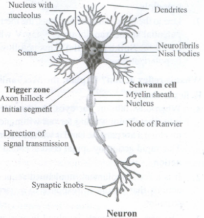
Draw a diagram of a neuron and label the parts.
14.
Describe the transport of respiratory gases.
15.
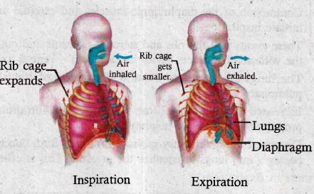
Compare the events in inspiration and expiration:
16.
17.
List the functions of the respiratory system.
18.
Explain the process of digestion in the stomach (OR)
What happens when food enters the stomach and explain the role of HCI in digestion process.
19.
Explain the process of physiology digestions of proteins.
20.
Describe the internal structure of human heart.
21.
Give a brief account on double circulation.
22.
Draw and label the mouthparts of the cockroach.
23.
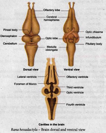
Explain the nervous system of frog.
24.
Write a note on three Domains? system of classification?
25.
Describe the digestive system of earthworm (Lampito).
1.
| S.No | Shark fish | Cat fish |
| 1. | It is a cat laginous fish and belongs to the class chondrichthyes | It is a bony fish and belongs to the class osteichthyes. |
| 2. | The skeleton is made of cartilage | The skeleton is made of calcified bones. |
| 3. | Upper Jaw of shark is not attached to the skill and moves independently. | Some bony fishes also have a second set of jaws (Pharyngeal Jaws) |
| 4. | The gill slits of shark are visible and not protected | The gills are covered by a bony plate. |
| 5. | Presence of third eyelid to protect the eye. | The eye has no protective coverings. |
2.
(i) Definition: The amount of blood pumped out by each ventricle per minute is called cardiac output (CO).
(ii) Heart rate (HR) and stroke volume (SV) are the products of CO.
(iii) HR or pulse is the number of beats per minute.
(iv) So pulse pressure = Systolic pressure - Diastolic pressure.
(v) SV is the volume of blood pumped out by one ventricle with each beat.
(vi) SV depends on ventricular systole/contraction.
(vii) CO = HR x SV
(viii) SV represents the difference between EDV (amount of blood that collects in a ventricle during diastole).
(ix) SV = EDV - ESV.
(x) Frank-Starling law of the heart, the critical factor controlling SV is the degree to which the cardiac myocytes are stretched just before they contract.
(xi) The most important factor stretching cardiac muscle is the amount of blood returning to the heart and distending its ventricles venous return.
(xii) SV performs the doubling of circulation during venous return during physical exercise.
(xiii) A balance between cardiac output and venous return is maintained by the pumping action of heart.
(xiv) As the heart serves as a double pump, each side can fail independently of the other.
(xv) If the left side of the heart fails. It results in pulmonary congestion and similarly the right side of the heart fails it results in the peripheral congestion.
(xvi) Frank-Starling effect protects the heart from abnormal increase in blood volume.
3.
Electrocaridogram (ECG): An electrocardiogram (ECG) records the electrical activity of the heart over a period of time using electrodes placed on the skin, arms, legs and chest. It records the changes in electrical potential across the heart during one cardiac cycle. A normal ECG shows 3 waves designated as P wave, QRS complex and T wave.
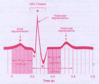
P Wave (atrial depolarisation): It is a small upward wave and indicates the depolarisation of the atria. This is the time taken for the excitation to spread through atria from SA node. Contraction of both atria lasts for around 0.8 - 1.0 sec.
PQ Interval (AV node delay): It is the onset of P wave to the onset of QRS complex. This is from the start of depolarisation of the atria to the beginning of ventricular depolarisation. It is the time taken for the impulse to travel from the atria to the ventricles (0.12 - 0.21 see). It is the measure of AV conduction time.
QRS Complex (ventricular depolarisation): No separate wave for atrial depolarisation in the ECG is visible. Atrial depolarisation occurs simultaneously with the ventricular depolarisation. The normal QRS complex lasts for 0.06-0.09 sec. QRS complex is shorter than the P wave, because depolarisation spreads through the Purkinjie fibres. Prolonged QRS wave indicates delayed conduction through the ventricle, often caused due to ventricular hypertrophy or due to a block in the branches of the bundle of His.
ST Segment: It lies between the QRS complex and T wave. It is the time during which all regions of the ventricles are completely depolarised and reflects the long plateau phase before repolarisation. In the heart muscle, the prolonged depolarisation is due to retardation of K+ efflux and is responsible for the plateau. The ST segment lasts for 0.09 sec.
T wave (ventricular depolarisation): It represents ventricular depolarisation. The duration of the T wave is longer than QRS complex because repolarisation takes place simultaneously throughout the ventricular depolarisation.
4.
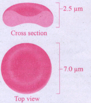
Red blood cells are abundant than the other blood cells. There are about 5 million to 5.5 millions of RBC mm-3 0f blood in a healthy man and 4.5-5.0 millions of RBC mm-3 in healthy women. The RBCs are very small with the diameter of about 7μm (micrometer). The structure of RBC is shown in Figure. The red colour of the RBC is due to the presence of a respiratory pigment, haemoglobin dissolved in the cytoplasm. Haemoglobin plays an important role in the transport of respiratory gases and facilitates the exchange of gases with the fluid outside the cell (tissue fluid). The biconcave shaped RBCs increases the surface area to volume ratio, hence oxygen diffuses quickly in and out of the cell. The RBCs are devoid of nucleus, mitochondria, ribosomes and endoplasmic reticulum. The absence of these organelles accommodates more haemoglobin thereby maximising the oxygen carrying capacity of the cell. The average life span of RBCs in a healthy individual is about 120 days after which they are destroyed in the spleen (graveyard / cemetery of RBCs) and the iron component returns to the bone marrow for reuse. Erythropoietin is a hormone secreted by the kidneys in response to low oxygen and helps in differentiation of stem cells of the bone marrow to erythrocytes (erythropoiesis) in adults. The ratio of red blood cells to blood plasma is expressed as Haematocrit.
5.
(i) Aves are commonly known as birds. The characteristic feature of Aves is the presence of feathers and the ability to fly except for flightless birds. eg. Ostrich, Kiwi.
(ii) The forelimbs are modified into wings, and the hind limbs are adapted for walking, running, swimming and perching.
(iii) The skin is dry and devoid of glands except the oil gland or preen gland at the base of the tail.
(iv) The exoskeleton consists of epidermal feathers, scales, claws on legs and the horny covering on the beak.
(v) The endoskeleton is fully ossified (bony) and the long bones are hollow with air cavities (pneumatic bones).
(vi) The pectoral muscles of flight (pectoralis major and pectoralis minor) are well developed.
(vii) Respiration is by compact, elastic, spongy lungs that are continuous with air sacs to supplement respiration.
(viii) The heart is four chambered.
(ix) Urinary bladder is absent.
(x) Sexes are separate with well marked sexual dimorphism.
(xi) All birds are oviparous. Eggs are megalecithal and cleidoic. Fertilization is internal.
(xii) eg. Corvus (Crow), Columba (Pigeon), Pavo (Peacock).
6.
(i) Amphibians are the first vertebrates and tetrapods to live both in aquatic as well as terrestrial habitats.
(ii) They are poikilothermic.
(iii) Their body is divisible into the head and trunk and most of them have two pairs of limbs; tail may or may not be present.
(iv) Their skin is smooth or rough, moist, pigmented and glandular.
(v) Eyes have eyelids and the tympanum represents the ear.
(vi) Respiration is by gills, lungs and through the skin.
(vii) Heart is three chambered.
(viii) Kidneys are mesonephric. Sexes are separate and fertilization is external.
(ix) They are oviparous and development is indirect. They show hibernation and aestivation.
(x) eg. Bufo (Toad), Rana (Frog).
7.
They are exclusively marine and are commonly called sea squirts, Mostly sessile, some pelagic or free swimming, exist as solitary and colonial forms. Body is unsegmented and covered by a test or tunic. Adult forms are sac like.. Coelom is absent, but has an atrial cavity surrounding the pharynx. Notochord is present only in the tail region of the larval stage, hence named urochordata. Alimentary canal is complete and circulatory system is of open type. The heart is ventral and tubular.
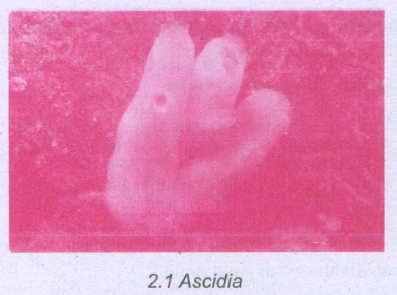
Respiration is through gill slits and clefts. Dorsal tubular nerve cord is present only in the larval stage and a single dorsal ganglion is present in the adults. Mostly hermaphrodites, development indirect and includes a free swimming tadpole larva with chordate characters. Retrogressive metamorphosis is seen. eg. Ascidia.
8.
(i)Annelids are the first segmented animals to evolve.
(ii) They are aquatic or terrestrial, free living but some are parasitic.
(iii) They are triploblastic, bilaterally symmetrical, schizocoelomates and exhibit organ system level of body organisation.
(iv) The coelom with coelomic fluid creates a hydrostatic skeleton and aids in locomotion.
(v) Their elongated body is metamerically segmented and the body surface is divided into segment or metameres.
(vi) Internally the segments are divided from one another by partitions called septa. This phenomenon is known as metamerism.
(vii) Aquatic annelids like Nereis have lateral appendages called parapodia, which help in swimming.
(viii) The circulatory system is of closed type and the respiratory pigments are hemoglobin and chlorocruorin.
(ix) Nervous system consists of paired ganglion connected by the lateral nerves to the double ventral nerve cord.
(x) They reproduce sexually. Development is direct or indirect and includes a trochophore larva. eg. Lampito mauritii (Earth worm).
9.
(i) The scientific name should be italicized in printed form and if handwritten, it should be underlined separately.
(ii) The generic name's (Genus) first alphabet should be in uppercase.
(iii) The specific name, (species) should be in lowercase.
(iv) The scientific names of any two organisms are not similar.
(v) The name or abbreviated name of the scientist who first publishes the scientific name mal be written after the species name along with the year of publication. ego Lion-Felis leo Linn., 1758 or Felis leo L., 1758.
(vi) If the species name is framed after any person's name the name of the species shall end with 'i', 'ii' or 'ae'. eg. A new species of a ground-dwelling lizard (Cyrtodactylus) has been discovered and named after scientist Varad Giri, Cyrtodactylus varadgirii.
10.
(i) It is a composite gland or mixed gland as it serves as both an exocrine gland in digestion and an endocrine gland in chemical co-ordination
(ii) It lies just below the stomach
(iii) It looks like a leaf
(iv) It is cream coloured
(v) It is made up of two major tissues like acini and islets of langerhans
(vi) Acini secretes the digestive enzymes
(vii) Islets of langerhans secrete hormones like insulin and glucagon
(viii) It has one to two million of islets of langerhans
(ix) In each islet there are
(i) 60% of islets of alpha cells
(ii) 25% of islets of beta cells
(iii) 10-15% of islets of delta cells
(iv) Alpha cells secrete glucagon
(v) Beta cells secrete insulin
(vi) Delta cells secrete somatostatin
Hormonal activities
a) Insulin
(i) A peptide hormone secreted by beta cells of islets of pancreas
(ii) Mainly regulates glucose homeostasis
(iii) Its main effect is to lower the blood glucose levels by increasing uptake of glucose by cells for oxidation
(iv) Eg: muscle and fat cells (glycolysis)
(v) It inhibits the breakdown of glycogen into glucose (glycogenolysis)
(vi) It prevents the conversion of proteins and fats into glucose (gluconeogenesis)
(vii) So it is called as hypoglycemic hormone
b) glucagon
(i) A Polypeptide hormone secreted by alpha cells of islets of pancreas
(ii) It is a potent hyperglycemic hormone
(iii) It acts on the liver and promotes glycogenesis, glycogenolysis and gluconeogenesis
(iv) It releases glucose from the liver cells / hepatocytes to increase the blood sugar level.
(v) Prolonged hyperglycemic condition by this hormone leads in diabetes mellitus.
11.
| S.No. | Sympathetic Neural System (SNS) | Parasympathetic Neural system (PNS) |
|---|---|---|
| 1. | SNS originates in the thoracic and lumbar region of the spinal cord | PNS originates in the cranial region of the brain and the sacral region of the spinal cord |
| 2. | Sympathetic ganglia are linked up to form a chain. | Its ganglia remain isolated. |
| 3. | Preganglionic fibres are short and the postganglionic fibres are long. | Preganglionic fibres are long and the postganglionic fibres are short. |
| 4. | Noradrenaline is produced at the terminal ends of the postganglionic fibres at the effector organs. Hence the system is adrenergic | Acetylcholine is produced at the terminal ends of the postganglionic fibres at the effector organs. Hence the system is cholinergic. |
| 5. | Active during stressful conditions preparing the body to face them. | Active during relaxing times restoring normal activity after a stress |
| 6. | The overall effect is excitatory and stimulating. | The overall effect is inhibitory. |
| 7. | It is considered as the flight or fight system. | It is considered as 'The Rest and Digest System' or 'The Feed and Breed System'. |
12.
Reflex action:
1. When dust falls in our eyes, the eyelids close I immediately not waiting for our willingness; on touching a hot pan, the hand is withdrawn rapidly. This rapid action by spinal cord is called reflex action.
2. It is a fast, involuntary, unplanned sequence of actions that occurs in response to a particular -stimulus.
3. The nervous elements involved in carrying out the reflex action constitute a reflex arc or in other words the pathway followed by a nerve impulse to produce a reflex action is called a reflex arc.
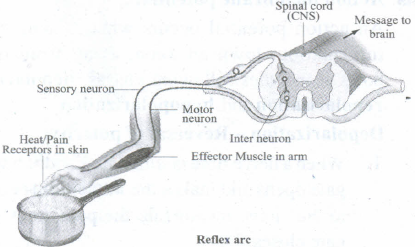
Functional components of a reflex arc:
1. Sensory Receptor - It is a sensory structure that responds to a specific stimulus.
2. Sensory Neuron - This neuron takes the sensory impulse to the grey (afferent) matter of the spinal cord through the dorsal root of the spinal cord.
3. Interneurons - One or two interneurons may serve to transmit the impulses from the sensory neuron to the motor neuron.
4. Motor Neuron - It transmits impulse from CNS to the effector organ.
5. Effector Organs - It may be a muscle or gland which responds to the impulse received.
13.
14.
(i) It is a transportary/chemical/blood respiration.
(ii) It occurs between alveoli and tissue fluid.
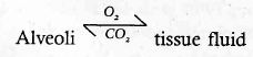
(iii) It mainly occurs in two steps
(A) Transport of O2
(B) Transport of CO2
(A) Transport of O2
(i) About 97% of O2 is transported by respiratory pigment haemoglobin which has more affinity with O2 to form HbO2 and poorly soluble in blood plasma.
(ii) HbO2 is formed firstly in the blood of pulmonary veins.
(iii) About 3% of O2 is transported by blood plasma under dissolved condition.
(iv) The rate at which haemoglobin binds with O2 is regulated by the pO2 (95 mm).
(v) Each haemoglobin contains four haem ions therefore four O₂ molecules are transported.
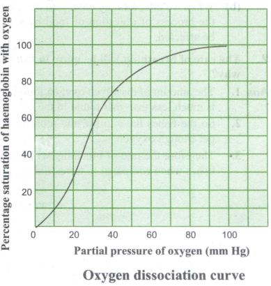
Oxygen dissociation curve
(i) In the alveoli pO2 is high (104 mm) and PCo2 is low (40 mm)
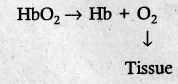
(ii) In the alveoli the temperature is low and concentration of H+ is also less. This conditions promote the formation of HbO₂ (Hb+ O₂→ HbO₂
(iii) In the tissues the pO2 is low (30 mm) and the pCO2 is high and high temperature promotes the dissociation of O2 from haemoglobin
(iv) Sigmoid (s-shaped) curve appears in oxygen dissociation curve. It is formed when % saturation of haemoglobin with O2 is plotted against pO2. This is called an oxygen haemoglobin dissociation curve.
(v) This curve appears in tissue and it is a steep slope for pO2 values between 10 and 50 mm Hg and then flattens between 70 and 100 mm Hg.
(vi) Under normal physiological state every 100 mL of oxygenated blood can deliver about 5 mL of O2 to the tissues.
(B) Transport of CO2
(i) CO2 is transported by
(a) Blood plasma: under dissolved state about 7-10% of CO2 is transported by plasma as CO2 is more soluble in blood plasma water.
(b) Bound with haemoglobin: about 20-25% of dissolved CO2 is bound and carried in the RBCs as HbCO2 (carbaminohaemoglobin).

(ii) Hb + CO2 occurs in tissues to form HbCO2
(iii) HbCO2 → Hb + CO2 occurs in alveoli of lungs
(C) As bicarbonate (HCO-3) in plasma about 70% of CO2 is transported as HCO-3 ions
(iv) The formation of HCO-3 ions is influenced by pCO2 and the degree of haemoglobin oxygenation with ferrous ions.
(v) RBCs contain a high concentration of the enzyme, carbonic anhydrase but a small quantity of this enzyme is also found in plasma of blood.
(vi) At the tissues the pCO2 is high due to catabolism and diffuses into blood to form HCO-3 and H+ ions.
(vii) The CO2 diffuses into RBCs and forms HbCO3 and into plasma water to form H2CO3 (carbonic acid).
(viii) H2CO3 is unstable and it is catalysed by carbonic anhydrase.
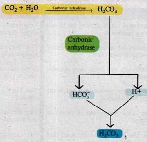
(ix) The HCO-3 moves quickly from the RBCs into plasma. From here it is transported to the lungs.
(x) At the alveolar site where pCO2 is low, the reaction is reversed leading to the formation of CO2 and H2O.
(xi) Thus CO2 trapped as HCO-3 at the tissue level is transported to the alveoli and released out as CO2.
(xii) Every 100 mL of deoxygenated blood delivers 4 mL of CO2 to the alveoli for elimination.
15.
| Inspiration | Expiration |
| Respiratory centre initiates the stimuli during inspiration \(\downarrow\) Impulses are carried to the inspiratory muscles through nerves. \(\downarrow\) Diaphragm and inspiratory muscles contract \(\downarrow\) The intrapulmonary pressure is reduced \(\downarrow\) The alveolar pressure increases than the atmospheric pressure. \(\downarrow\) Air flows into the alveoli until the alveolar pressure equalizes the atmospheric pressure and the alveoli get inflated |
Respiratory centre terminates the stimuli during expiration \(\downarrow\) The diaphragm and inspiratory muscles relax \(\downarrow\) Chest wall contracts and the thoracic volume gets reduced. \(\downarrow\) The intrapulmonary pressure is reduced. \(\downarrow\) The alveolar pressure increases than the atmospheric pressure. \(\downarrow\) Air is sent out due to the contraction of alveoli \(\downarrow\) Air flows into the alveoli until the alveolar pressure equalizes the atmospheric pressure and the alveoli get inflated |
16.

17.
The five primary functions of the respiratory system are
1. To exchange O2 and CO2 between the atmosphere and the blood.
2. To maintain homeostatic regulation of body pH.
3. To protect us from inhaled pathogens and pollutants.
4. To maintain the vocal cords for normal communication (vocalization).
5. To remove the heat produced during cellular respiration through breathing.
18.
Digestion in the stomach:
1. Food remains in the stomach for 4 to 5 hours, the rhythmic peristaltic movement churns and mixes the food with gastric juice and make it into a creamy liquid called chyme.
2. The gastric secretion is partly controlled by autonomic reflexes.
3. The secretion of gastric juice begins when the food is in the mouth.
4. The gastric juice contains HCI and proenzymes. The proenzyme pepsinogen, on exposure to HCI gets converted into the active enzyme pepsin which converts proteins into proteoses and peptones (peptides).
5. The HCI provides an acidic medium (PH1.8) which is optimum for pepsin, kills bacteria and other harmful organisms and avoids putrefaction.
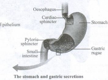
6. The mucus and bicarbonates present in the gastric juice play an important role in lubrication and protection of the mucosal epithelium from the eroding nature of the highly acidic HCI.
7. Another proteolytic enzyme found in gastric juice of infants is rennin helps in the digestion of milk protein, caseinogen to casein in the presence of calcium ions. This enzyme secretion gradually reduces with aging.
19.
(a) Stomach
(i) The acidic medium of gastric juice (1.8 pH) mainly aids in the digestion of proteins
(ii) The digestion of proteins starts in the stomach.
(iii) After a meal gastric artery supplies more oxygenated blood for the stomach for the digestion of proteins.
(iv) HCI secreted by oxyntic cells activates the pepsinogen and rennin for the digestion of proteins.
(v) Inactive pepsinogen and inactive rennin are the precursors of active enzymes found in gastric juice secreted by parietal cells
Enzymatic reactions
(i) Proteins + pepsinogen + HCl → pepsin
(ii) Pepsin proteins → polypeptides + tri, dipeptides + amino acids
(iii) Rennin + HCl + milk protein casein → curd
(b) Duodenum
(i) Pancreatic juice containing inactive enzymes like chymotrypsinogen and trypsinogen and active carboxyl peptidase involves in the digestion of proteins
(ii) The catalytic enzyme converts in active enzyme chymotrypsinogen into active chymotrypsin and inactive, trypsinogen into active trypsin
Enzymatic reactions:
(i) Chymotrypsin + proteins → polypeptides + tri, dipeptides and amino acids
(ii) Trypsin + protein →polypeptides + tri, di peptides and amino acids
(iii) Carboxyl peptidase + polypeptides + tri, di peptides → free amino acids
(c) Small intestine
(i) Intestinal juice or succus entericus contains an enzyme peptidase
Enzymatic reaction
Dipeptides, Tripeptides + Peptidase → Free amino acids
20.
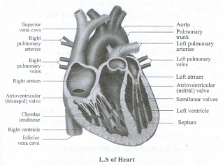
Structure of human heart (L.S)
(i) Raymond de viessens in 1706 described the structure of heart.
(ii) Heart is made up of cardiac muscles.
(iii) Heart lies in the thoracic cavity (mediastinum) the space between the two lungs).
(iv) Its apex part is slightly tilted towards left.
(v) In adult human it weighs about 300g.
(vi) Its size is roughly equal to a closed fist of fore limb.
(vii) Heart is divisible into 4 chambers upper two small atria and lower two large ventricles.
(viii) Atria are separated by interauricular septum.
(ix) Ventricles are separated by inter ventricular septum.
(x) Due to the presence of these septae mixing of blood is prevented.
(xi) The walls of ventricles are thicker than atria.
(xi) The thickness of ventricles is due to the presence of papillary muscles.
(xii) The heart is enveloped by three layers an outer epicardium, middle myocardium and an inner endocardium.
(xiii) Over all the heart is enveloped by double membranous peritoneum called pericardium.
(xiv) The space between the two membranes is inter pericardial space filled by pericardial fluid.
(xv) Atria communicate with the ventricles by atrio ventricular apertures.
(i) Such apertures are guarded by atrio ventricular valves
(a) Tricuspid valve between right atrium and right ventricle
(b) Bicuspid or mitral valve between left atrium and left ventricle
(ii) Atrio-ventricular valves prevent the backward flow of blood from ventricles to atria.
(iii) Semi lunar valves in ventricles prevent the backward flow of blood into ventricles and direct the flow of blood into aorta and pulmonary artery in the left ventricle and right ventricle respectively.
(iv) Each semilunar valve is made up of three half moon shaped cusps.
(v) The middle myocardium shows an irregular muscular ridges called trabeculae corneae.
(vi) The modified trabeculae corneae are called chordae tendineae.
(vii) The chordae tendineae hold the valves in the heart under fixed position.
(viii) The chordae tendineae are attached to the lower end of the heart by papillary muscles.
(ix) The main venous blood vessels like Inferior vena cava and superior vena cava and a small coronary sinus collect deoxygenated blood from entire body and transport such deoxygenated into right atrium.
(x) Pulmonary veins from the lungs bring oxygenated to the left atrium.
21.
(i) It was firstly described by William Harvey in 1628.
(ii) In higher terrestrial vertebrates two types of circulation occur because blood enters the heart twice.
(a) First type of circulation (systemic)
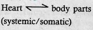
(i) Aorta arises from left ventricle comes out of the heart and forms arterial system to supply O₂, nutrients and other organics and inorganics to the tissues
(ii) Similarly the venous system collects CO2, NH3 and other excreta to right atrium of the heart
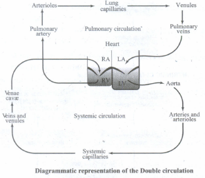
(b) 2nd type of circulation (Pulmonary)
(i) It occurs between heart and lungs.
(ii) It brings both decarboxylation and oxygenation.
(iii) Pulmonary artery arises from right atrium divides into right and left branches to enter the lungs.
(iv) Oxygenation and decarboxylation are performed in the alveoli of lungs.
(v) A paired pulmonary veins that arise from the lungs bring the oxygenated blood to left atrium of the heart.
(vi) Veins that enter the heart (right atrium) have decreased pressure than that of arteries found closer to heart.
(vii) A completion of cardiac cycle needs double circulation in higher animals like mammals.
22.
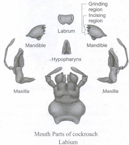
23.
Divisions of nervous system
1. Central Nervous System (CNS)
2. Peripheral Nervous System (PNS)
3. Autonomic Nervous System (ANS)
(i) CNS consists of brain and spinal cord
(ii) PNS consists of cranial nerves and spinal nerves
(iii) ANS consists of sympathetic and parasympathetic nervous systems
(iv) CNS consists of 10 paired cranial nerves and 10 paired spinal nerves
(v) controls visceral involuntary activities.
(vi) Brain lies in cranial cavity.
(vii) Brain and spinal cord are covered by two meninges.
(viii) Such two meninges are an outer piamater and an inner duramater
(ix) Brain is divisible into
(i) Fore brain (Prosencephalon).
(ii) Mid brain (Mesencephalon).
(iii) Hind brain (Rhombencephalon).
(i) Fore brain:
(i) It is the anterior most and largest part consisting of a paired olfactory lobes, cerebral hemispheres and a diencephalon.
(ii) Anterior part of the olfactory lobe is narrow and free but is fused posteriorly
(iii) Olfactory lobes aid in the sense of smell.
(iv) The cavity of olfactory lobes is called as ventricle.
(v) Cerebral hemispheres (telencephalon) are the largest part of fore brain.
(ii) Mid brain:
(i) It includes two large, oval shaped optic lobes having cavities and are called as optic ventricles.
(iii) Hind brain:
(i) It consists of the cerebellum and medulla oblongata.
(ii) Cerebellum balances the body in movements and maintains posture of the body.
(iii) Cerebellum is narrow, thin transverse band followed by medulla oblongata.
(iv) Medulla oblongata passes out through the foramen magnum then it continues as spinal cord.
(v) The spinal cord passes through the vertebral column.
24.
(i) Definition: The system emphasizes the separation of prokaryotes into two domains, Bacteria and Archaea and 3rd Eukarya.
(ii) Carl woese (1977) divided Domains into three types. They are
a. Domain Archaea
b. Domain Bacteria and
c. Domain Eukarya
a) Domain Archaea:
Eg: Single celled organisms (prokaryotes)
(i) They live under extreme climatic conditions
(ii) As they have the ability to grow under extreme conditions like volcano vents, hot springs and polar ice caps, the prokaryotes are called as extremophiles
(iii) Chemosynthetic bacteria can synthesize food by the oxidation of inorganics like hydrogen sulphide (H₂S)
(iv) Methane producing organisms are methanogens
(v) Organisms living in salty environment are halophiles
(vi) Organisms live under higher temperatures are thermoacidophiles.
b) Domain Bacteria:
(i) Members are prokaryotes like bacteria
(ii) Cells have no definite true nucleus
(iii) DNA is large, circular, coiled, doubled stranded and naked
(iv) Histones are absent
(v) No membrane bound organelles
(vi) Sub-organelles like 70s type of ribosomes are membranous
(vii) Cell wall is made up of peptidoglycans
(viii) Many bacteria are decomposers (eat on dead decaying organisms)
(ix) Some are photosynthetic and pathogenic
(x) The beneficial probiotic and harmful pathogenic bacteria are diversely populated
(xi) Blue-green algae (prokaryotic plants) like cyanobacteria are photosynthetic they release O2
(xii) Such bacteria balance atmospheric O2 level and these were previously anaerobic and become aerobic during the advancement of changes of geographical periods.
c) Domain Eukarya:
(i) All animals belong to Eukarya
(ii) True nucleus and membrane bound organelles and sub-organelles are found-
(iii) DNA in the nucleus is arranged as a linear chromosome
(iv) Histones proteins are present
(v) Ribosomes are of 80s type but 70s type of ribosomes are found in chloroplasts and mitochondria (like in prokaryotes)
(vi) Animals in this domain are classified into Protista, Fungi, Plantae and Animalia
25.
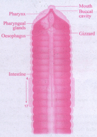
Digestive system consists of Alimentary canal and digestive glands
(A) Alimentary canal
(i) It is straight passing through out the length of the body from mouth to pygidium
(ii) Mouth lies in 1st and 2nd true segments and opens into buccal cavity
(iii) Buccal cavity opens posteriorly into a thick muscular pharynx at 3rd and 4th segments
(iv) Pharynx is surrounded by the pharyngeal glands
(v) A small narrow tube oesophagus lying in the 5th segment continues into muscular gizzard lying in 6th segment
(vi) Gizzards grinds the soil rich in humus (decaying leaves)
(vii) A narrow small intestine extends from 7th to 14th segments where digestion and absorption take place, and its dorsal wall is folded to form typhlosole consisting of blood vessels and it also increases the digestive and absorptive areas to complete the digestion
(viii) A sacculated large intestine extends between 15th to last segment functions in excretion and opens exterior by anus or pygidium to eliminate excreta

11th Standard Syllabus & Materials
11th Standard
TN 11th Tamil பீடு பெற நில் - செய்யுள் - காவடிச்சிந்து Important Questions And Answers Study Material - QB365 Set A
NEW11th Standard
TN 11th Tamil பீடு பெற நில் - உரைநடை - மலை இடப்பெயர்கள் : ஓர் ஆய்வு Important Questions And Answers Study Material - QB365 Set A
NEW11th Standard
TN 11th Tamil மாமழை போற்றுதும் - துணைப்பாடம் - யானை டாக்டர் Important Questions And Answers Study Material - QB365 Set A
NEW11th Standard
TN 11th Tamil மாமழை போற்றுதும் - செய்யுள் - ஐங்குறுநூறு Important Questions And Answers Study Material - QB365 Set A



Tamilnadu Stateboard 11th Standard Subjects

Maths

Commerce

Economics
Biology

Business Maths and Statistics

Accountancy

Computer Science

Physics

Chemistry
Maths
Biology
Economics
Physics
Chemistry

History
Business Maths and Statistics

Computer Science
Accountancy

Computer Applications
History
Computer Technology
Commerce
Computer Applications
Computer Technology

Tamil

English

French
Tamilnadu Stateboard Standards