 100% FREE ACCESS
11th Standard Syllabus & Materials
11th Standard
TN 11th Tamil இயற்கை வேளாண்மை,சுற்றுச்சூழல் -செய்யுள் - மனோன்மணீயம் Important Questions And Answers Study Material - QB365 Set A
NEW11th Standard
TN 11th Tamil என்னுயிர் என்பேன் -துணைப்பாடம் - இசைத்தமிழர் இருவர் Important Questions And Answers Study Material - QB365 Set A
NEW11th Standard
TN 11th Tamil மொழி கலை -செய்யுள் - ஒவ்வொரு புல்லையும் Important Questions And Answers Study Material - QB365 Set A
NEW11th Standard
TN 11th Tamil பீடு பெற நில் - இலக்கணம் - பகுபத உறுப்புகள் Important Questions And Answers Study Material - QB365 Set A
NEW11th Standard
TN 11th Tamil பீடு பெற நில் - துணைப்பாடம் - வாடிவாசல் Important Questions And Answers Study Material - QB365 Set A
NEW11th Standard
TN 11th Tamil பீடு பெற நில் - செய்யுள் - குறுந்தொகை Important Questions And Answers Study Material - QB365 Set A
Published on: 13/05/2022
QB365 provides detailed and simple solution for every Creative Questions in class 11 Biology Subject. It will helps to get more idea about question pattern in every Creative questions with solution.
latest Creative QuestionsDownload Tamil Nadu 11th Standard Biology question papers, model tests, one-mark questions, important questions, and public exam papers in PDF format. Free study materials and answer keys for TN State Board students.
Questions + Answers key
Take MCQ Biology Test1.
Explain the importance of blood pressure.
2.
Give a brief account on cardiac cycle.
3.
Write a short note on the life cycle of earthworm.
4.
Explain the digestion in the stomach.
5.
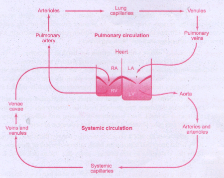
Show the double circulation by way of diagrammatic sketch.
6.
What are the general features of Reptilians?
7.
How are the vertebrates are further divided by divisions? Write the differences between them.
8.
What are the fundamental distinct features of all chordates?
9.
Write an account on common characters of Arthropoda.
10.
Describe the morphology of earthworms with neat diagram.
11.
How are reflex actions classified?
12.
Explain Depolarization in the conduction of nerve impulse.
13.
Write a note on the structure of contractile protein.
14.
Write note on
a) Emphysema and
b) Tuberculosis.
15.
Explain Transport of carbon dioxide during respiration.
16.
Write a note on surfactants.
17.
Explain the structure of the liver with diagram. Add a note on its functions.
18.
Explain the process of physiology of digestion of fats.
19.
Write a note on disorders of the circulatory system (Any two).
20.
Explain coagulation of blood.
21.
Describe the digestive system of cockroach.
22.
23.
Describe the female reproductive system of cockroach.
24.
Write a note on Hemichordata.
25.
Give a brief account on Ctenophora.
1.
Blood pressure is the pressure exerted on the surface of blood vessels by the blood. This pressure circulates the blood through arteries, veins, and capillaries. There are two types of pressure, the systolic pressure, and the diastolic pressure. Systolic pressure is the pressure in the arteries as the chambers of the heart contracts. Diastolic pressure is the pressure in the arteries when the heart chambers relax. Blood pressure is measured using a sphygmomanometer (BP apparatus). It is expressed as systolic pressure I diastolic pressure. Normal blood pressure in man is about 120/80mm Hg. Mean arterial pressure is a function of cardiac output and resistance in the arterioles. The primary reflex pathway for homeostatic control of mean arterial pressure is the baroreceptor reflex. The baroreceptor reflex functions every morning when you get out of bed. When you are lying flat the gravitational force is evenly distributed. When you stand up, gravity causes blood to pool in the lower extremities. The decrease in blood pressure upon standing is known as orthostatic hypotension. Orthostatic reflex normally triggers baroreceptor reflex. This results in increased cardiac output and increased peripheral resistance which together increases the mean arterial pressure.
2.
Definition: The events that occur at the beginning of heart beat and lasts until the beginning of next heart beat is called as cardiac cycle.
(i) It's duration is 0.8 sec.
(ii) It includes 5 phases.
Phase: 1
(i) It is a ventricular systole.
(ii) Auricular pressure is high.
(iii) Ventricular pressure is low.
(iv) A.V valves open.
(v) Semilunar valves are closed.
(vi) Blood flows from atria to ventricles.
Phase: 2
(i) It is an atrial systole.
(ii) Atria contract and ventricles relax.
(iii) Ventricles are filled by blood until EDV (End Diastolic Volume).
(iv) EDV is related to the length of cardiac muscle fibre.
(v) More the muscles are stretched, greater than EDV and SV.
Phase: 3
(i) It is a ventricular systole.
(ii) It occurs due to isovolumetric contraction.
(iii) AV valves are forced by the force of the ventricular contraction.
(iv) Ventricles show increased pressure.
(v) There is no change in the size of the muscle fibre length.
Phase: 4
(i) It is a ventricular systole or ventricular ejection.
(ii) Without backward flow of blood.
(iii)This point is the end of systolic volume (ESV).
(iv) Blood is pumped into aortic valves and pulmonary valves of left ventricle and right ventricle respectively.
Phase: 5
(i) It is a ventricular diastole.
(ii) Relaxation of ventricles starts.
(iii) Pressure is high in arteries and low in ventricles.
(iv) Semi lunar valves are closed.
(v) The heart returns to phase-1 of the cardiac cycle.
3.
Lampito mauritii begins its life cycle, from the fertilized eggs. The eggs are held in a protective cocoon. These cocoons have an incubation period of about 14- 18 days after which they hatch to release juveniles (Figure). The juveniles undergo changes into nonclitellate forms in phase - I after about 15 days, which then develops a clitellum, called the clitellate at the end of the growth phase - II taking 15 - 17 days to complete. During the reproductive stage, earthworms copulate, and later shed their cocoons in the soil after about 10 days. The life cycle of Lampito mauritii takes about 60 days to complete.
4.
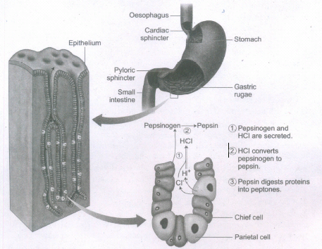
Food remains in the stomach for 4 to 5 hours, the rhythmic peristaltic movement churns and mixes the food with gastric juice and make it into a creamy liquid called chyme. The gastric secretion is partly controlled by autonomic reflexes. The secretion of gastric juice begins when the food is in the mouth. The gastric juice contains HCI and proenzymes. The proenzyme pepsinogen, on exposure to HCI gets converted into the active enzyme pepsin which converts proteins into proteoses and peptones (peptides). The HCI provides an acidic medium (pH 1.8) which is optimum for pepsin, kills bacteria and other harmful organisms and avoids putrifaction. The mucus and bicarbonates present in the gastric juice play an important role in lubrication and protection of the mucosal epithelium from the eroding nature of the highly acidic HCI (Figure). Another proteolytic enzyme found in gastric juice of infants is rennin which helps in the digestion of milk protein, caseinogen to casein in the presence of \ calcium ions. This enzyme secretion gradually reduces with aging.
5.
6.
(i) They are mostly terrestrial animals and their body is covered by dry, and cornified skin with epidermal scales or scutes.
(ii) Reptiles have three chambered heart but four chambered in crocodiles.
(iii) All are cold blooded amniotes.
(iv) Most reptiles lay cleidoic eggs with extraembryonic membranes like amnion, allantois, chorion and yolk sac.
(v) Excretion by metanephric kidneys and are uricotelic.
(vi) They are monoecious. Internal fertilization takes place and all are oviparous.
(vii) eg. Calotes (Garden Lizard), Draco (Flying Lizard), Crocodilus (Crocodile).
7.
(i) Subphylum Vertebrata is divided into two divisions, Agnatha and Gnathostomata.
(ii) Agnatha includes jawless fish-like aquatic. vertebrates without paired appendages. Notochord persists in the adult.
(iii) Agnatha includes one important class-Cyclostomata.
(iv) Gnathostomata includes jawed vertebrates with paired appendages. Notochord is replaced partly or wholly by the vertebral column.
(v) Gnathostomata includes jawed fishes (Pisces) and Tetrapoda (amphibia, reptilia, aves and mammals). The superclass Pisces includes all fishes which are essentially aquatic forms with paired fins for swimming and gills for respiration. Pisces includes cartilaginous fishes (Chondrichthyes) and bony fishes (Osteicthyes).
8.
All chordates possess three fundamental distinct features at some stage of their life cycle, they are:
(i) Presence of elongated rod like notochord below the nerve cord and above the alimentary canal. It serves as a primitive internal skeleton. It may persist throughout life in lancelets and lampreys. In adult vertebrates, it may be partially or completely replaced by backbone or vertebral column.
(ii) A dorsal hollow or tubular fluid filled nerve cord lies above the notochord and below the dorsal body wall. It serves to integrate and co-ordinate the body functions. In higher chordates, the anterior end of the nerve cord gets enlarged to form the brain and the posterior part becomes the spinal cord, protected inside the vertebral column.
(iii) Presence of pharyngeal gill slits or clefts in all chordates at some stage of their life cycle. It is a series of gill slits or clefts that perforates the walls of pharynx and appears during the development of every chordate. In aquatic forms, pharyngeal gill slits are vascular, lamellar and form the gills for respiration. In terrestrial chordates, traces of non-functional gill clefts appear during embryonic developmental stages and disappear later. Besides the above said features, chordates are bilaterally symmetrical, triploblastic, coelomates with organ system level of organisation; they possess post anal tail, closed circulatory system with a ventral myogenic heart except in Amphioxus.
9.
(i) This is the largest phylum of the Kingdom Animalia and includes the largest class called Insecta.
(ii) They are bilaterally symmetrical, segmented, triploblastic and schizocoelomate animals with organ system grade of body organisation.
(iii) They have jointed appendages which are used for locomotion feeding and are sensory in function.
(iv) Body is covered by chitinous exoskeleton for protection and to prevent water loss, It is shed off periodically by a process called moulting or ecdysis.
(v) The body consists of a head, thorax, and abdomen with a body cavity called haemocoel.
(vi) Respiratory organs are gills, book gills, book lungs or trachea.
(vii) Circulatory system is of open type.
(viii) Sensory organs like antennae, eyes (compound and simple), statocysts (organs of balance I equilibrium) are present.
(ix) Excretion takes place through malpighian tubules, green glands, coxal glands, etc.
(x) They are mostly dioecious and oviparous; fertilization is usually internal.
(xi) Life history includes many larval stages followed by metamorphosis. eg. Limulus.
10.
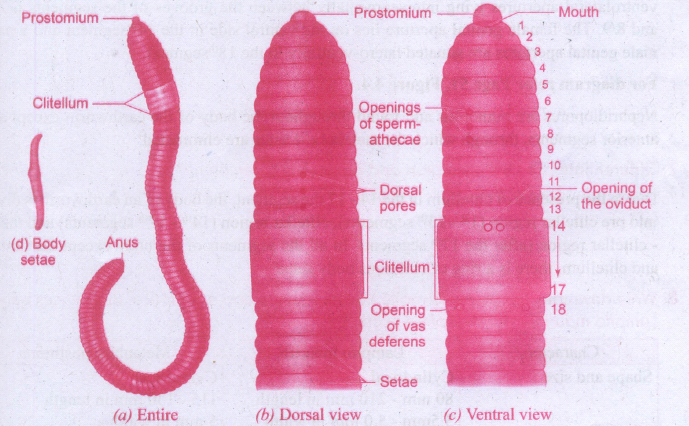
Lampito mauritii is commonly found in Tamil Nadu. It has a long and cylindrical narrow body which is bilaterally symmetrical. L. mauritii is 80 to 210 mm in length with a diameter of 3.5 - 5 mm, and is light brown in colour, with purplish tinge at the anterior end. This colour of the earthworm is mainly due to the presence of porphyrin pigment. The body of the earthworm is encircled by a large number of grooves which divides it into a number of compartments called segments or metameres. L. mauritii consists of about 165 - 190 segments. The dorsal surface of the body is marked by a dark mid dorsal line (dorsal blood vessel) along the longitudinal axis of the body. The ventral surface is distinguished by the presence of genital openings. The mouth is found in the centre of the first segment of the body, called the peristomium. Overhanging the mouth is a small flap called the upper lip or prostomium. The last segment has the anus called the pygidium. In mature worms, segments 14 to 17 may be found swollen with a glandular thickening of the skin called the clitellum. This helps in the formation of the cocoon. Due to the presence of clitellum, the body of an earthworm is divided into pre clitellar region (1st- 13th segments), clitellar region (14th - 17th segments) and the post - clitellar region (after the 17th segment). In all the segments of the body except the first,last and clitelhim, there is a ring of chitinous body setae. This body setae arises from a setigerous sac of the skin and it is curved as S - shaped. Setae can be protruded or retracted and their principal role is in locomotion.
11.
Reflex action:
1. When dust falls in our eyes, the eyelids close immedi~tely not waiting for our willingness; on touching a hot pan, the hand is withdrawn rapidly. This rapid action by spinal cord is called reflex action.
2. It is a fast, involuntary, unplanned sequence of actions that occurs in response to a particular stimulus.
3. The nervous elements involved in carrying out the reflex action constitute a reflex arc or in other words the pathway followed by a nerve impulse to produce a reflex action is called a reflex arc.
There are two types of reflexes. They are
1. Unconditional reflex is an inborn reflex for an unconditioned stimulus. It does not need any past experience, knowledge or training to occur; Eg: Blinking of an eye when a dust particle about to fall into it, sneezing and coughing due to foreign particle entering the nose or larynx.
2. Conditioned reflex is a response to a stimulus that has been acquired by learning. This does not naturally exists in animals. Only an experience makes it a part of the behaviour. Eg: Excitement of salivary gland on seeing and smelling a food. The conditioned reflex was first demonstrated by the Russian physiologist Pavlov in his classical conditioning experiment in a dog. The cerebral cortex controls the conditioned reflex.
12.
Action membrane potential:
An action potential occurs when a neuron sends information down an axon, away from the cell body. It includes following phases, depolarization, repolarisation and hypopolarization
Depolarization - Reversal of polarity:
1. When a nerve fiber is stimulated, sodium voltage gate opens and makes the axolemma permeable to Na+ ions; meanwhile, the potassium voltage gate closes.
2. As a result, the rate of flow of Na+ ions into the axoplasm exceeds the rate of flow of K+ ions to the outside fluid [ECF].
3. Therefore, the axolemma becomes positively charged inside and negatively charged outside. This reversal of electrical charge is called Depolarization.
4. During depolarization, when enough Na+ ions enter the cell, the action potential reaches a certain level, called threshold potential [-55mV].
5. The particular stimulus which is able to bring the membrane potential to the threshold is called threshold stimulus.
6. The action potential occurs in response to a threshold stimulus but does not occur at subthreshold stimuli. This is called all or none principle.
7. Due to the rapid influx of Na+ ions, the membrane potential shoots rapidly up to +45m V which is called the Spike potential. This is followed by Repolarization.
13.
1. Contraction of the muscle depends on the presence of contractile proteins such as actin and myosin.
2. The thick filament of muscle fibres are composed of the protein myosin. The thin filaments are formed of three types of proteins called actin, tropomyosin and troponin. These four proteins are known as contractile proteins.
Thick myofilament:
3. Each myosin filament is made up of a monomer called meromyosin which has two regions, a globular head, a short arm called heavy meromyosin (HMM) and a tail called light meromyosin (LMM).
The globular head is an ATPase enzyme which has binding sites for ATP and actin - binding site.
Thin myofilament:
4. It is composed of two intertwined actin molecules. Actin has polypeptide subunits called G-actin (globular actin) and F - actin (Filamentous actin). F- actin is an polymer of monomeric G-Actin and also has a myosin - binding site.
5. The thin filaments also contain several regulatory proteins like tropomyosin and troponin which help in regulating muscles contraction.
14.
(a) Emphysema:
1. Emphysema is chronic breathlessness caused by the gradual breakdown of the thin walls of the alveoli decreasing the total surface area of a gaseous exchange. i.e., widening of the alveoli .is called emphysema.
2. The major cause of this disease is cigarette smoking, which reduces the respiratory surface of the alveolar walls.
(b) Tuberculosis:
1. It is a respiratory disorder caused by Mycobacterium tuberculosis.
2. This infection mainly occurs in the lungs and bones.
3. Collection of fluid between the lungs and the chest wall is the main complication of this disease.
15.
Transport of Carbon dioxide: Blood transports CO2 from the tissue cells to the lungs in three ways,
(i) Dissolved in plasma: About 7-10% of CO2 is transported in a dissolved form in the plasma.
(ii) Bound to haemoglobin: About 20-25% of dissolved CO2 is bound and carried in the RBCs as carbaminohaemoglobin (HbCO2)
CO2 + Hb \(\leftrightarrows \) Hb CO2
(iii) As bicarbonate ions in plasma: About 70% of CO2 is transported as bicarbonate ions.
1. This is influenced by pCO2 and the degree of haemoglobin oxygenation. RBCs contain a high concentration of the enzyme, carbonic anhydrase, Whereas small amounts of carbonic anhydrase is present in the plasma.
2. At the tissues the pCO2 is high due to catabolism and diffuses into the blood to form HCO3-and H+ ions. When CO2 diffuses into the RBCs, it combines with water forming carbonic acid (H2CO3) catalyzed by carbonic anhydrase. Carbonic acid is unstable and dissociates into hydrogen and bicarbonate ions.
3. Carbonic anhydrase facilitates the reaction in both directions.
CO2 + H2O  H2CO3
H2CO3
HCO3- + H+
4. The HCO3- moves quickly from the RBCs into the plasma, where it is carried to the lungs. At the alveolar site where pCO2 is low, the reaction is reversed leading to the formation of CO2 and water. Thus CO2 trapped as HCO3- at the tissue level it is transported to the alveoli and released out as CO2.
5. Every 100mL of deoxygenated blood delivers 4mL of CO2 to the alveoli for elimination.
16.
1. The thin squamous epithelial cells of the alveoli are composed of Type I and Type II cells.
2. Type I cells are very thin so that gases can diffuse rapidly through them.
3. Type II cells are thicker, synthesize and secrete a substance called Surfactant.
4. Surfactants are the thin non-cellular films made of protein and phospholipids covering the alveolar membrane.
5. The surfactant lowers the surface tension in the alveoli and prevents the lungs from collapsing.
6. It also prevents pulmonary oedema.
7. Premature Babies have low levels of surfactant in the alveoli may develop the newborn respiratory distress syndrome (NRDS) because the synthesis of surfactants begins only after the 25th week of gestation.
17.
Structure of the Liver:
1. The liver, the largest gland in our body is situated in the upper right side of the abdominal cavity, just below the diaphragm.
2. The liver consists of two major left and right lobes; and two minor lobes. These lobes are connected with the diaphragm. Each lobe has many hepatic lobules (functional unit of the liver) and is covered by a thin connective tissue sheath called the Glisson's capsule.
3. Liver cells (hepatic cells) secrete bile which is stored and concentrated in a thin muscular sac called the gallbladder.
4. The duct of gallbladder (cystic duct) along with the hepatic duct from the liver forms the common bile duct. The bile duct passes downwards and joins with the main pancreatic duct to form a common duct called hepato-pancreatic duct.
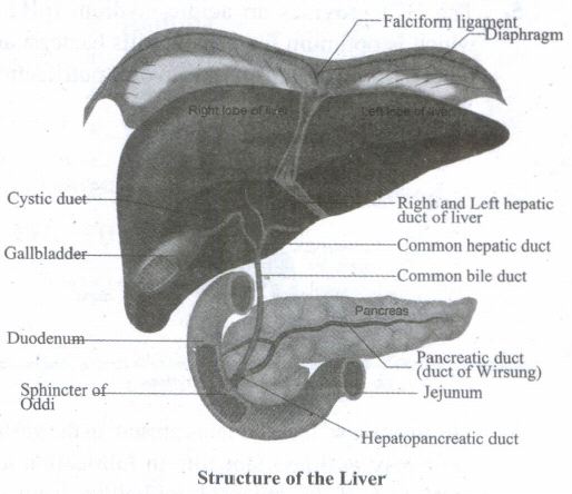
5. The opening of the hepato-pancreatic duct into' the duodenum is guarded by a sphincter called the sphincter of Oddi. The liver has the high power of regeneration and liver cells are replaced by new ones every 3-4 weeks.
Apart from bile secretion, the liver also performs several functions. They are
1. Destroys ageing and defective blood cells.
2. Stores glucose in the form of glycogen or disperses glucose into the bloodstream with the help of pancreatic hormones.
3. Stores of fat-soluble vitamins and iron.
4. Detoxifies toxic substances.
5. Involves in the synthesis of non- essential amino acids and urea
18.
(a) Stomach
(i) Weak lipase found in gastric juice emulsifies and saponifies fats in the presence of HCI.
(b) Duodenum of small intestine
(i) Pancreatic lipase acts on fats or emulsified and saponified fats in duodenum
Enzyme reaction
Lipase + fats → Di and mono glycerides
(c) Jejunum of small intestine
(i) Intestinal juice or succus entericus contains lipases enzymes.
Enzymatic reaction
Diglycerides and Monoglycerides \(\underrightarrow { Lipases } \) Fatty acids + Glycerol
19.
Hypertension:
1. It is the most common circulatory disease. The normal blood pressure in man is 120/80 mmHg.
2. In cases when the diastolic pressure exceeds 90 mm Hg and the systolic pressure exceeds 150 mm Hg persistently, the condition is called hypertension.
3. Uncontrolled hypertension may damage the heart, brain and kidneys.
Coronary heart disease:
1. It occurs when the arteries are lined by atheroma. The build-up of atheroma contains cholesterol, fibres, dead muscle and platelets and is termed Atherosclerosis.
2. The cholesterol-rich atheroma forms plaques in the inner lining of the arteries making them less elastic and reduces the blood flow.
3. Plaque grows within the artery and tends to form blood clots, forming coronary thrombus. Thrombus in a coronary artery results in heart attack.
20.
Coagulation of blood:
The mechanism by which excessive blood loss is prevented by the formation of clot is called blood coagulation or clotting of blood. Schematic representation of blood coagulation is shown below.
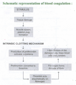
1. The clotting process begins when the endothelium of the blood vessel is damaged and the connective tissue in its wall is exposed to the blood.
2. Platelets adhere to collagen fibres in the connective tissue and release substances that form the platelet plug which provides emergency protection against blood loss.
3. Clotting factors released from the clumped platelets or damaged cells mix- with clotting factors in the plasma. The protein called prothrombin is converted to its active form called thrombin in the presence of calcium and vitamin K.
4. Thrombin helps in the conversion of fibrinogen to fibrin threads. The threads of fibrin become interlinked into a patch that traps blood cell and seals the injured vessel until the wound is healed.
5. After sometime fibrin fibrils contract, squeezing out a straw-coloured fluid through a meshwork called serum.
21.
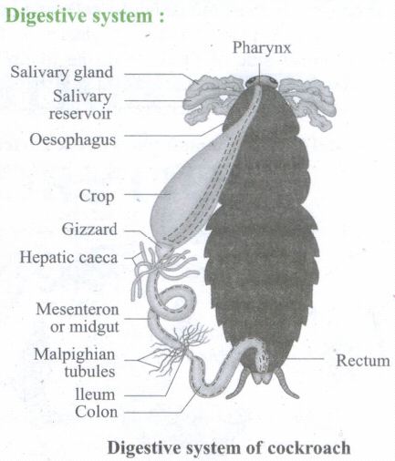
1. The digestive system of cockroach consists of
(a) Alimentary canal and
(b) Digestive glands.
2. The alimentary canal is present in the body cavity and is divided into three regions:
(a) Foregut
(b) Midgut and mesenteron and
(c) Hindgut
(a) Foregut:
(i) It includes pre- oral cavity, mouth, pharynx and oesophagus.
(ii) It also includes salivary glands and mouth parts.
(iii) Oral cavity or cibarium opens into oesophagus through pharynx.
(iv) Oesophagus opens into a sac like structure called the crop.
(v) The crop is followed by gizzard or proventriculus.
(vi) Gizzard has an outer layer of thick circular muscles and thick inner cuticle forming six highly chitinous plates called teeth.
(vii) Gizzard aids in the grinding of food particles.
(b) Mid gut
(i) It is a short, narrow tube found behind the gizzard.
(ii) It is glandular in nature.
(iii) At the junctional region of the gizzard are eight like tubular blind processes called the hepatic caecae or enteric caecae which function as liver in higher chordates.
(iv) Digestion and absorption of digested food are performed.
(c) Hind gut
(i) At the junction of midgut and hind gut 100-150 yellow coloured malpighian tubules are present which are excretory in function.
(ii) Hind gut is broader than the midgut.
(iii) Hind gut includes ileum, colon, and rectum.
(iv) Rectum opens exterior by anus.
(v) Salivary glands, the glandular cells and hepatic caecae are the digestive glands.
(vi) A paired salivary glands are found one on either side of the crop of foregut in the thorax.
(vii) The glandular cells of midgut and hepatic caecae of foregut secrete digestive juice.
22.
23.
1. The female reproductive system of cockroach consists of a pair of ovaries, vagina, genital pouch, colleterial glands, spermathecae and the external genitalia.
2. A pair of ovaries lies laterally in the 2nd and 6th abdominal segment.
3. Each ovary is formed of a group of eight ovarian tubules or ovarioles, containing a chain of developing ova.
4. The lateral oviducts of each ovary unite into a broad median common oviduct known as vagina, which opens into the genital chamber.
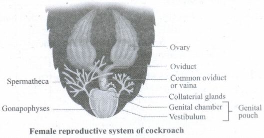
5. The vertical opening of the vagina is the female genital pore.
6. A pair of spermathecae is present in the 6th segment, which opens by a median aperture in the dorsal wall of the genital pouch.
7. During copulation, the ova descend to the genital chamber, where they are fertilized by the sperms.
8. A pair of white and branched colleterial glands present behind the ovaries forms a hard egg case called Ootheca around the eggs. Genital pouch is formed by the 7th 8th and 9th abdominal sterna.
9. The genital pouch has two chambers, (a) genital chamber into which the vagina opens and (b) oothecal chamber where oothecae are formed.
10. Three pairs of plate like chitinous structures called gonapophyses are present around the female genital aperture. These gonapophyses guide the ova into the ootheca as ovipositors.
24.
1. Hemichordates were earlier treated as a subphylum of Chordata (or Prochordata). They are now regarded to be an independent phylum of invertebrates, close to Echinodermata. The animals of this group possess the characters of invertebrates as well as chordates.
2. This phylum consists of a small group of wormlike, soft marine animals, mostly tubiculous and commonly called the 'acorn worms' or 'tongue worms'.
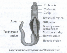
3. They are bilaterally symmetrical, triploblastic and coelomate animals with organ system level of organisation.
4. Their body is cylindrical and is divided into three regions, the anterior proboscis, a short collar and a long trunk.
5. Most hemichordates are ciliary feeders. Their circulatory system is simple and open or lacune type with a dorsal heart.
6. Respiration is through paired gill slits opening into the pharynx.
7. Excretion is by a single proboscis gland or glomerulus situated in the proboscis.
8. Nervous system is primitive.
9. Sexes are separate and exhibit sexual mode of reproduction; Fertilization is external. Development is indirect with a free swimming tornaria larva. Eg:Balanoglossus, Saccoglossus.
25.
(i) Animals exclusively live in marine.
(ii) Animals are radially symmetrical diploblastic leading in tissue grade of organization.
(iii) The middle layer between ecto and endodermal layers called mesoglea.
(iv) Consisting of amoebocytes and smooth muscle cells.
(v) Eight external rowed ciliated comb plates aid in locomotion.
(vi) Animals emit light (biolumuniscence).
(vii) Animals are commonly called as sea walnuts or Comb jellies.
(viii) No nematocysts
(ix) Lasso cells or colloblasts are found to capture food.
(x) Digestion is both intra and inter cellular.
(xi) Animals are bisexual (monoeceous).
(xii) Fertilization is external.
(xiii) The larva is cydippid
Eg: Pleurobrachia and Ctenophore.

11th Standard Syllabus & Materials
11th Standard
TN 11th Tamil பீடு பெற நில் - செய்யுள் - காவடிச்சிந்து Important Questions And Answers Study Material - QB365 Set A
NEW11th Standard
TN 11th Tamil பீடு பெற நில் - உரைநடை - மலை இடப்பெயர்கள் : ஓர் ஆய்வு Important Questions And Answers Study Material - QB365 Set A
NEW11th Standard
TN 11th Tamil மாமழை போற்றுதும் - துணைப்பாடம் - யானை டாக்டர் Important Questions And Answers Study Material - QB365 Set A
NEW11th Standard
TN 11th Tamil மாமழை போற்றுதும் - செய்யுள் - ஐங்குறுநூறு Important Questions And Answers Study Material - QB365 Set A



Tamilnadu Stateboard 11th Standard Subjects

Maths

Commerce

Economics
Biology

Business Maths and Statistics

Accountancy

Computer Science

Physics

Chemistry
Maths
Biology
Economics
Physics
Chemistry

History
Business Maths and Statistics

Computer Science
Accountancy

Computer Applications
History
Computer Technology
Commerce
Computer Applications
Computer Technology

Tamil

English

French
Tamilnadu Stateboard Standards