 100% FREE ACCESS
11th Standard Syllabus & Materials
11th Standard
TN 11th Tamil இயற்கை வேளாண்மை,சுற்றுச்சூழல் -செய்யுள் - மனோன்மணீயம் Important Questions And Answers Study Material - QB365 Set A
NEW11th Standard
TN 11th Tamil என்னுயிர் என்பேன் -துணைப்பாடம் - இசைத்தமிழர் இருவர் Important Questions And Answers Study Material - QB365 Set A
NEW11th Standard
TN 11th Tamil மொழி கலை -செய்யுள் - ஒவ்வொரு புல்லையும் Important Questions And Answers Study Material - QB365 Set A
NEW11th Standard
TN 11th Tamil பீடு பெற நில் - இலக்கணம் - பகுபத உறுப்புகள் Important Questions And Answers Study Material - QB365 Set A
NEW11th Standard
TN 11th Tamil பீடு பெற நில் - துணைப்பாடம் - வாடிவாசல் Important Questions And Answers Study Material - QB365 Set A
NEW11th Standard
TN 11th Tamil பீடு பெற நில் - செய்யுள் - குறுந்தொகை Important Questions And Answers Study Material - QB365 Set A
Published on: 08/06/2021
QB365 provides detailed and simple solution for every book back questions in class 11 Biology subject.It will helps to get more idea about question pattern in every book back questions with solution.
Download Tamil Nadu 11th Standard Biology question papers, model tests, one-mark questions, important questions, and public exam papers in PDF format. Free study materials and answer keys for TN State Board students.
Questions + Answers key
Take MCQ Biology Test1.
2.
Explain the sliding- filament theory of muscle contraction.
3.
What are the bones that make the pelvic girdle?
4.
What are the different types of rib bones that form the rib cage?
5.
List the disorders of the muscular system.
1.
2.
Mechanism of muscle contraction
(i) Sliding filament theory in 1954, Andrew F. Huxley and Rolf Niedergerke proposed the sliding-filament theory to explain muscle contraction.
(ii) According to this theory, overlapping actin and myosin filaments of fixed length slide past one another in an energy requiring process, resulting in muscle contraction.
(iii) The contraction of muscle fibre is a remarkable process that helps in creating a force to move or to resist a load.
(iv) The force which is created by the contracting muscle is called muscle tension.
(v) The load is a weight or force that opposes contraction of a muscle.
(vi) Contraction is the creation of tension in the muscle which is an active process and relaxation is the release of tension created by contraction.
(vii) Muscle contraction is initiated by a nerve impulse sent by the central nervous system (CNS) through a motor neuron.
(viii) The junction between the motor neuron and the sarcolemma of the muscle fibre is called the neuromuscular junction or motor end plate.
(ix) When nerve impulse reaches a neuromuscular junction, acetylcholine is released.
(x) It initiates the opening of multiple gated channels in sarcolemma. The action potential travels along the T-tubules and triggers the release of calcium ions from the sarcoplasmic reticulum.
(xi) The released calcium ions bind to troponin on thin filaments. The tropomyosin uncovers the myosin-binding sites on thin filaments.
(xii) Now the active sites are exposed to the heads of myosin to form a cross-bridge. During cross-bridge formation actin and myosin form a protein complex called actomyosin.
(xiii) Utilizing the energy released from hydrolysis of ATP, the myosin head rotates until it forms a 90° angle with the long axis of the filament.
(xiv) In this position myosin binds to an actin and activates a contraction relaxation cycle which is followed by a power stroke.
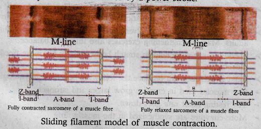
(xv) The power stroke (cross-bridge tilting) begins after the myosin head and hinge region tilt from a 90° angle to a 45° angle. The cross-bridge transforms into strong, high-force bond which allows the myosin head to swivel. When the myosin head swivels it pulls the attached actin filament towards the centre of the A-band.
(xvi) The myosin returns back to its relaxed state and releases ADP and phosphate ion. A new ATP molecule then binds to the head of the myosin and the cross-bridge is broken.
(xvii) At the end of each power stroke, each myosin head detaches from actin, then swivels back and binds to a new actin molecule to start another contraction cycle. This movement is similar to the motion of an oar on a boat.
(xviii) At the end of each power stroke, each myosin head detaches from actin, then swivels back and binds to a new actin molecule to start another contraction cycle.
(xix) The power stroke repeats many times until a muscle fibre contracts. The myosin heads bind, push and release actin molecules over and over as the thin filaments move toward the centre of the sarcomere.
(xx) The repeated formation of cross-bridge cycles cause the sliding of the filaments only but there is no change in the lengths of either the thick or thin filaments.
(xxi) The Z-discs attached to the actin filaments are also pulled inwards from both the sides, causing the shortening of the sarcomere (i.e. contraction). This process continues as long as the muscle receives the stimuli and with a steady flow of calcium ions.
(xxii) When motor impulse stops, the calcium ions are pumped back into the sarcoplasmic reticulum, results in the masking of the active sites of the actin filaments. The myosin head fails to bind with the active sites of actin and these changes cause the return of Z-discs back to their original position, i.e. relaxation.
3.
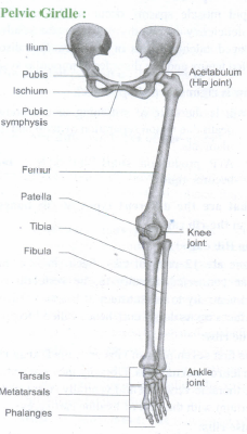
1. The pelvic girdle is a heavy structure specialized for weight bearing. It is composed of two hip bones called coxal bones that secure the lower limbs to the axial skeleton. Together, with the sacrum and coccyx, the hip bones form the basin-like bony pelvis.
2. Each coxal bone consists of three fused bones, ilium, ischium and pubis. At the point of fusion of ilium, ischium, and pubis a deep hemispherical socket called the acetabulum is present on the lateral surface of the pelvis.
3. It receives the head of the femur or thigh bone at the hip joint and helps in the articulation of the femur.
4. The ilium is the superior flaring portion of the hip bone. Each ilium forms a secure joint with the sacrum posteriorly. The ischium is a curved bar of bone. The V-shaped pubic bones articulate anteriorly at the pubic symphysis.
The Lower limb:
1. The lower limb consists of 30 bones which carries the entire weight of the erect body.
2. The bones of the lower limbs are thicker and stronger than the upper limbs. The three segments of each lower limb are the thigh, the leg or the shank and the foot.
3. The femur is the single bone of the thigh. It is the largest, longest and strongest bone in the body. The head of femur articulates with the acetabulum of the pelvis to form the hip joint.
4. Two parallel bones, the tibia and fibula, form the skeleton of the shank. A thick, triangular patella forms the kneecap, which protects the knee joint anteriorly and improves the leverage of thigh muscles acting across the knee.
5. The foot includes the bones of ankle, the tarsus, the metatarsus and the phalanges or toe bones. The foot supports our body weight and acts as a lever to propel the body forward while walking and running.
6. The tarsus is made up of seven bones called tarsals. The metatarsus consists of five bones called metatarsals. The arrangement of the metatarsals is parallel to each other. There are 14 phalanges in the toes which are smaller than those of the fingers.
4.
The Rib cage:
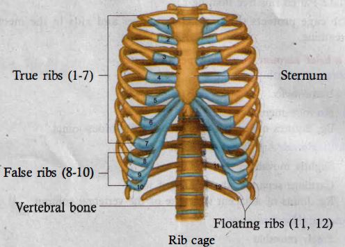
(i) There are 12 pairs of ribs.
(ii) Each rib is a thin flat bone connected dorsally to the vertebral column and ventrally to the sternum.
(iii) It has two articulation surfaces on its dorsal end hence called bicephalic.
(iv) The first seven pairs of ribs are called 'true ribs' or vertebro-sternal ribs.
(v) Dorsally they are attached to the thoracic vertebrae and ventrally connected to the sternum with the help of hyaline cartilages.
(vi) The 8th, 9th and 10th pairs of ribs do not articulate directly with the sternum but joined with the cartilaginous (hyaline cartilage) part of the seventh rib.
(vii) These are called 'false ribs' or vertebrochondral ribs.
(viii) The last 11th and 12th pairs of ribs are not connected ventrally. Therefore, they are called as 'floating ribs' or vertebral ribs.
(ix) Thoracic vertebrae, ribs and sternum together form the ribcage.
5.
Myasthenia gravis, Tetany, Muscle pull, Muscle Fatigue, Atrophy, Dystrophy.

11th Standard Syllabus & Materials
11th Standard
TN 11th Tamil பீடு பெற நில் - செய்யுள் - காவடிச்சிந்து Important Questions And Answers Study Material - QB365 Set A
NEW11th Standard
TN 11th Tamil பீடு பெற நில் - உரைநடை - மலை இடப்பெயர்கள் : ஓர் ஆய்வு Important Questions And Answers Study Material - QB365 Set A
NEW11th Standard
TN 11th Tamil மாமழை போற்றுதும் - துணைப்பாடம் - யானை டாக்டர் Important Questions And Answers Study Material - QB365 Set A
NEW11th Standard
TN 11th Tamil மாமழை போற்றுதும் - செய்யுள் - ஐங்குறுநூறு Important Questions And Answers Study Material - QB365 Set A



Tamilnadu Stateboard 11th Standard Subjects

Maths

Commerce

Economics
Biology

Business Maths and Statistics

Accountancy

Computer Science

Physics

Chemistry
Maths
Biology
Economics
Physics
Chemistry

History
Business Maths and Statistics

Computer Science
Accountancy

Computer Applications
History
Computer Technology
Commerce
Computer Applications
Computer Technology

Tamil

English

French
Tamilnadu Stateboard Standards